Lezcano Lisette Blanco, Alberti Amador Esteban, González Fraguela María Elena, Zaldívar Lelo de Larrea Guadalupe, Serrano Rosa Martha Pérez, Jiménez Luna Nadia Angélica, Camejo Rodríguez Dianet, Serrano Sánchez Teresa, Francis Turner Liliana, Estupiñán Díaz Bárbara, Vega Hurtado Yamilé, Fernández Jiménez Isabel
Experimental Neurophysiology Department. International Center of Neurological Restoration (CIREN) Ave. 25 No. 15805 e/158 and 160, Playa, Havana 10300, Cuba.
Faculty of Medicine, Autonomous University of Queretaro, Clavel Street No. 200, Col. Prados de la Capilla, Santiago de Querétaro, Querétaro 76176, Mexico.
Behav Sci (Basel). 2020 Oct 13;10(10):156. doi: 10.3390/bs10100156.
Neurotoxic lesion of the pedunculopontine nucleus (PPN) is known to cause subtle motor dysfunctions. However, motor coordination during advance on a discontinuous and elevated surface has not been studied. It is also not known whether there are changes in the mRNA expression of nuclear factor (erythroid-derived 2)-like 2 () in nigral tissue.
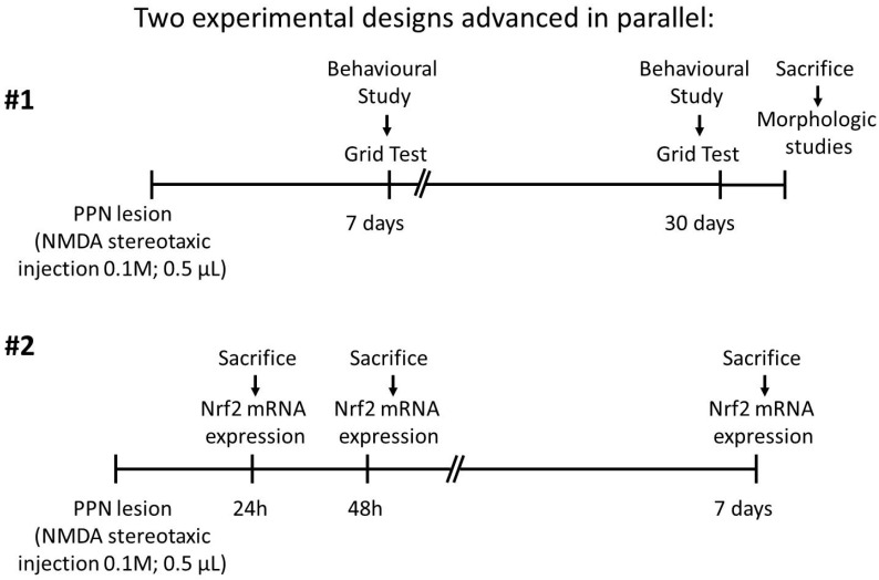
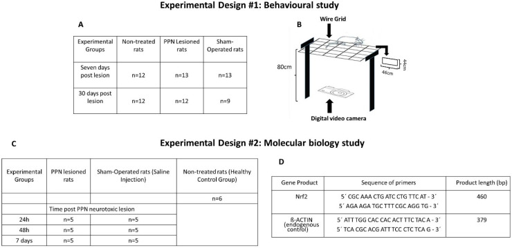
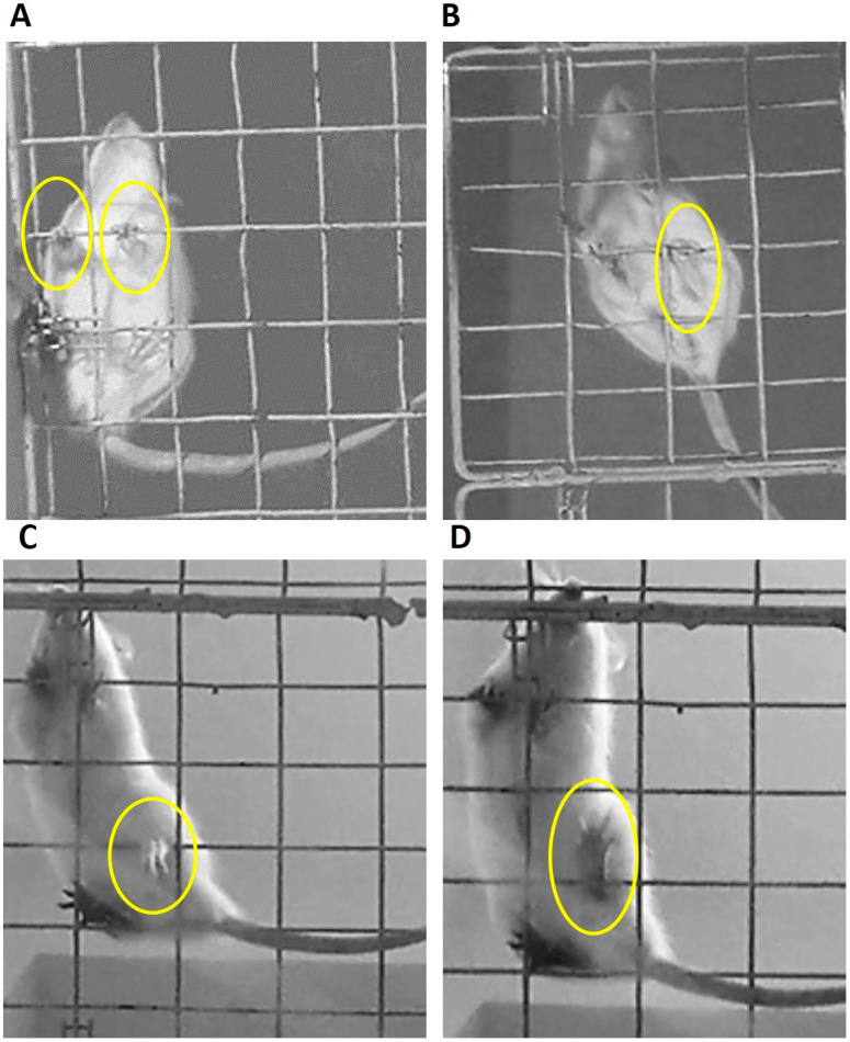
The effects of the unilateral neurotoxic lesion of the PPN in motor coordination evaluated through grid test and mRNA expression in nigral tissue were evaluated. Two experimental designs (ED) were organized: ED#1 behavioral study (7 and 30 days after PPN lesion) and ED#2 molecular biology study (24 h, 48 h and 7 days) after PPN lesion.
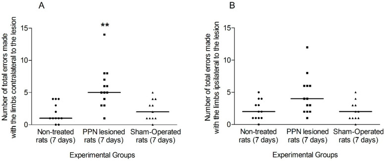
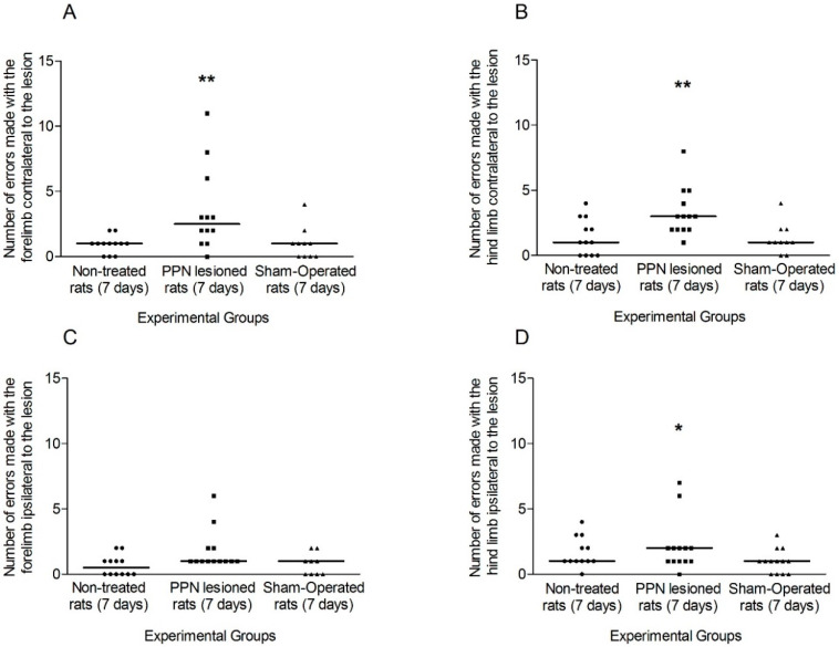
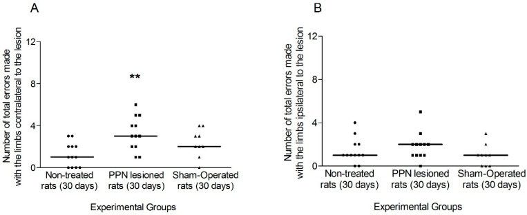
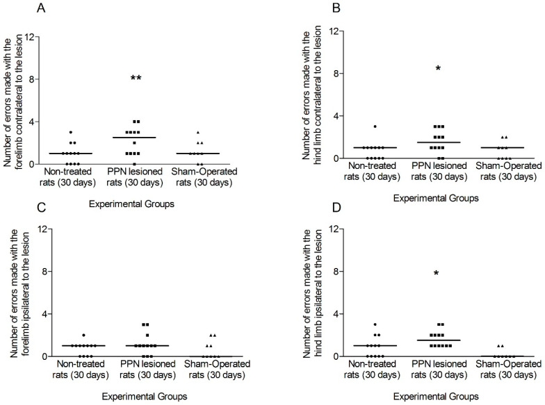
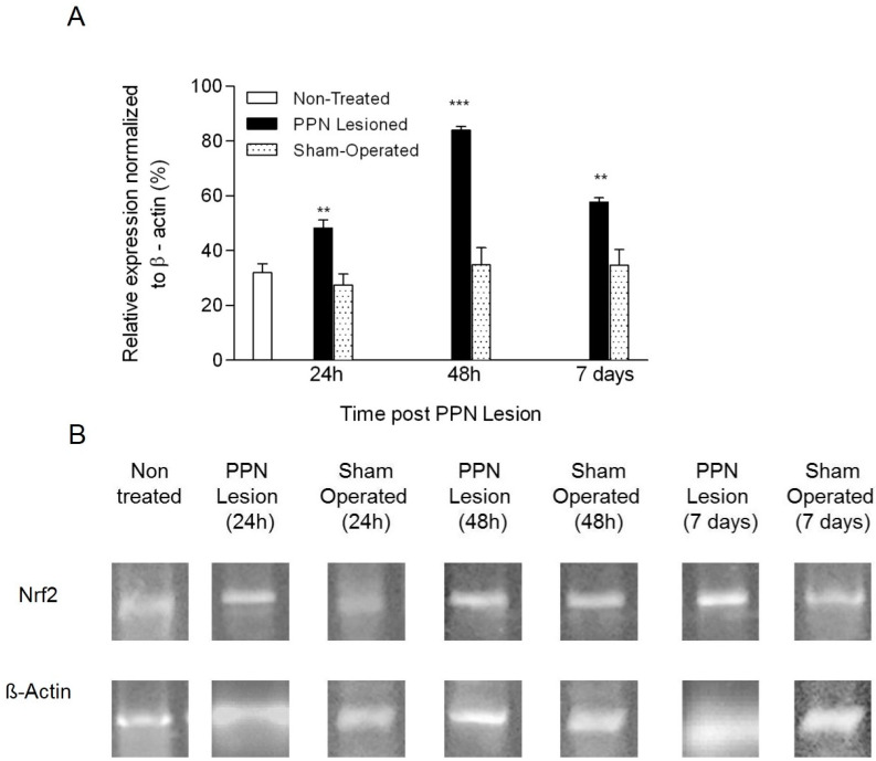
ED#1-The number of faults made with left limbs, were significant higher in the lesioned groups ( < 0.01) both 7 and 30 days post-lesion. The number of failures made by the right limbs, was also significantly higher ( < 0.05) vs. control groups. ED#2- mRNA expression showed an increase 24 h after PPN injury ( < 0.01), followed by a peak of expression 48 h post injury ( < 0.001).
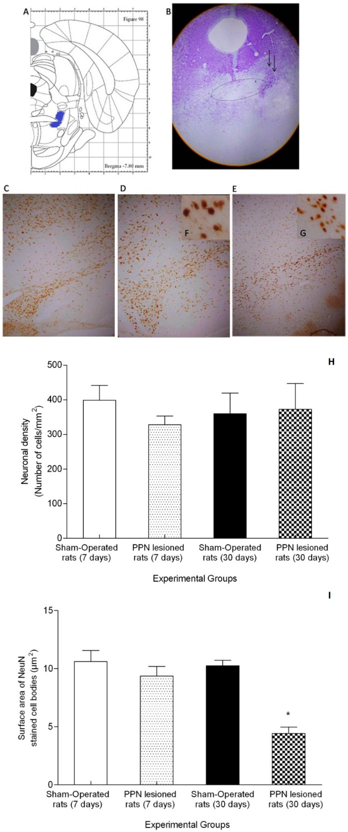
Disorders of motor coordination associated with PPN injury are bilateral. The increased mRNA expression could represent an adaptive response to oxidative stress in the nigral tissue following pontine injury.
已知脑桥脚核(PPN)的神经毒性损伤会导致细微的运动功能障碍。然而,在不连续且升高的表面上前进时的运动协调性尚未得到研究。黑质组织中核因子(红系衍生2)样2()的mRNA表达是否存在变化也不清楚。
通过网格试验评估PPN单侧神经毒性损伤对运动协调性的影响,并评估黑质组织中的mRNA表达。组织了两个实验设计(ED):ED#1行为学研究(PPN损伤后7天和30天)和ED#2分子生物学研究(PPN损伤后24小时、48小时和7天)。
ED#1-损伤组左肢出现的错误数量在损伤后7天和30天均显著高于对照组(<0.01)。右肢出现的失误数量与对照组相比也显著更高(<0.05)。ED#2-mRNA表达在PPN损伤后24小时增加(<0.01),随后在损伤后48小时达到表达峰值(<0.001)。
与PPN损伤相关的运动协调性障碍是双侧的。mRNA表达增加可能代表脑桥损伤后黑质组织对氧化应激的适应性反应。