Zhang Yongxiang, Li Yuechuan, Ye Zhen, Ma Hui
Department of Respiratory and Critical Care Medicine, Tianjin Chest Hospital, Tianjin, China (mainland).
Med Sci Monit. 2020 Oct 18;26:e925278. doi: 10.12659/MSM.925278.
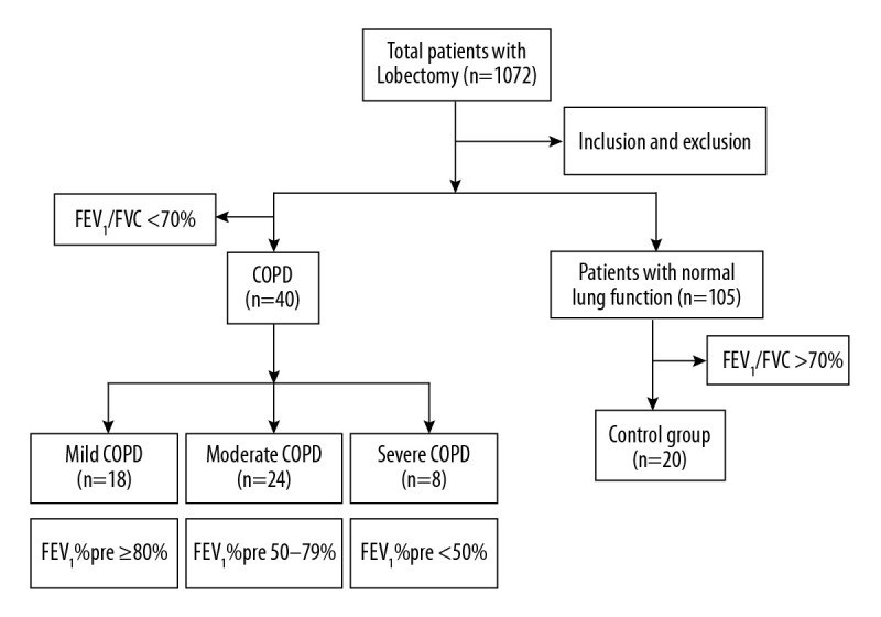
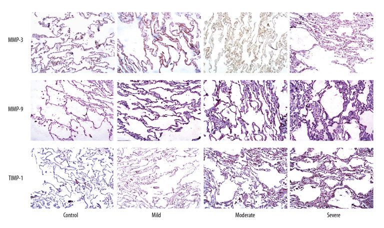
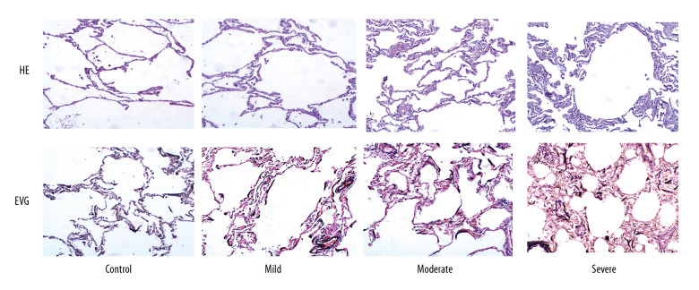
BACKGROUND This study investigated the relationship between the pathological alteration of alveolar septa and (1) pulmonary function and (2) matrix metalloproteinase (MMP)-2, MMP-9, and tissue inhibitor matrix metalloproteinase 1 (TIMP-1) expression in chronic obstructive pulmonary disease (COPD). MATERIAL AND METHODS Sixty patients with pulmonary disease were divided into control (n=20) and COPD (n=40) groups. Postoperative lung tissue specimens were examined. Hematoxylin and eosin and elastin van Gieson staining detected pathological alterations of pulmonary alveolar septa. Septa thickness was measured. MMP-2, MMP-9, and TIMP-1 expression levels were detected by immunohistochemical staining. Correlations were determined by Pearson analysis. RESULTS Forced expiratory volume in 1 s (FEV₁), forced vital capacity, FEV₁ percent predicted (FEV₁%pre), and diffusion capacity of carbon monoxide percent predicted (DLCO%pre) in COPD patients were significantly lower than in those of the control group (P<0.05). MMP-2, MMP-9, and TIMP-1 expression levels were significantly higher in the COPD group than in control, especially the severe group (P<0.05). Septa thickness was negatively correlated with FEV₁%pre (r=-0.335; P<0.05) and positively correlated with MMP-2 and TIMP-1 expression (P<0.05). Proportion of collagenous fiber was negatively correlated with FEV₁%pre and DLCO%pre (P<0.01), and positively correlated with MMP-2, MMP-9, and TIMP-1 expression (P<0.01). Proportion of elastic fibers was negatively correlated with collagenous fiber. CONCLUSIONS The pathological alteration of alveolar septa was correlated with pulmonary function and expression levels of MMP-2, MMP-9, and TIMP-1, which can play vital roles in COPD progression.
背景 本研究调查了慢性阻塞性肺疾病(COPD)中肺泡间隔的病理改变与(1)肺功能以及(2)基质金属蛋白酶(MMP)-2、MMP-9和基质金属蛋白酶组织抑制剂1(TIMP-1)表达之间的关系。材料与方法 将60例肺部疾病患者分为对照组(n = 20)和COPD组(n = 40)。对术后肺组织标本进行检查。苏木精-伊红染色和弹性蛋白-范吉森染色检测肺泡间隔的病理改变。测量间隔厚度。通过免疫组织化学染色检测MMP-2、MMP-9和TIMP-1的表达水平。采用Pearson分析确定相关性。结果 COPD患者的第1秒用力呼气容积(FEV₁)、用力肺活量、预测FEV₁百分比(FEV₁%pre)和预测一氧化碳弥散量百分比(DLCO%pre)显著低于对照组(P<0.05)。COPD组中MMP-2、MMP-9和TIMP-1的表达水平显著高于对照组,尤其是重度组(P<0.05)。间隔厚度与FEV₁%pre呈负相关(r = -0.335;P<0.05),与MMP-2和TIMP-1表达呈正相关(P<0.05)。胶原纤维比例与FEV₁%pre和DLCO%pre呈负相关(P<0.01),与MMP-2、MMP-9和TIMP-1表达呈正相关(P<0.01)。弹性纤维比例与胶原纤维呈负相关。结论 肺泡间隔的病理改变与肺功能以及MMP-2、MMP-9和TIMP-1的表达水平相关,它们在COPD进展中可能起重要作用。