Faraji-Bellée Carole-Anne, Cauliez Axelle, Salmon Benjamin, Fogel Olivier, Zhukouskaya Volha, Benoit Aurélie, Schinke Thorsten, Roux Christian, Linglart Agnès, Miceli-Richard Corinne, Chaussain Catherine, Briot Karine, Bardet Claire
Université de Paris, Laboratory Orofacial Pathologies, Imaging and Biotherapies UR 2496, Dental School, Montrouge, France.
APHP, Reference Center for Rare Disorders of the Calcium and Phosphate Metabolism, Dental Medicine Department, Bretonneau Hospital, Paris, France.
Front Cell Dev Biol. 2020 Sep 22;8:854. doi: 10.3389/fcell.2020.00854. eCollection 2020.
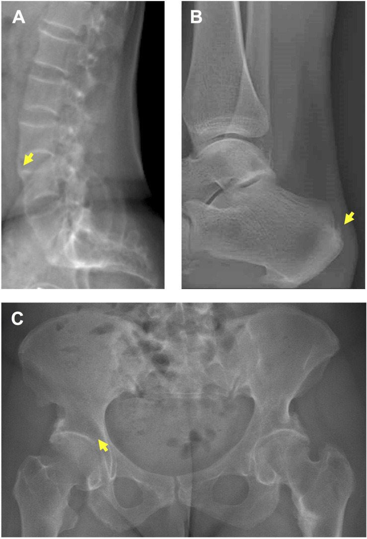
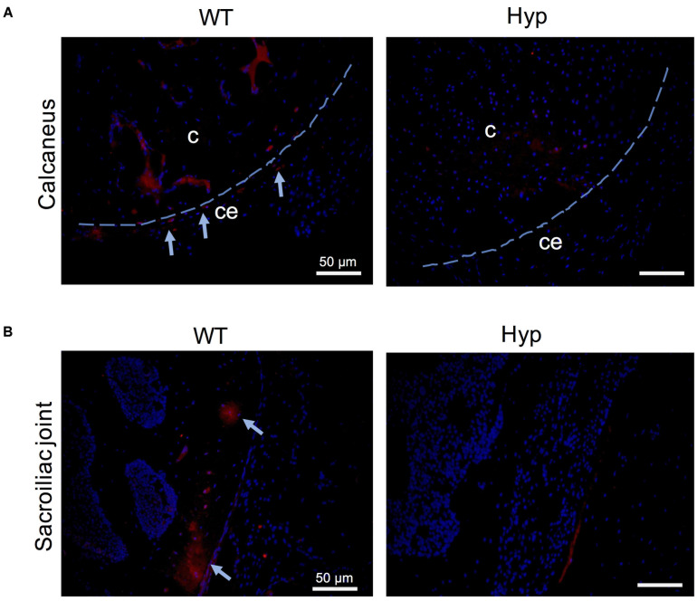
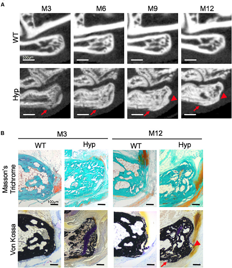
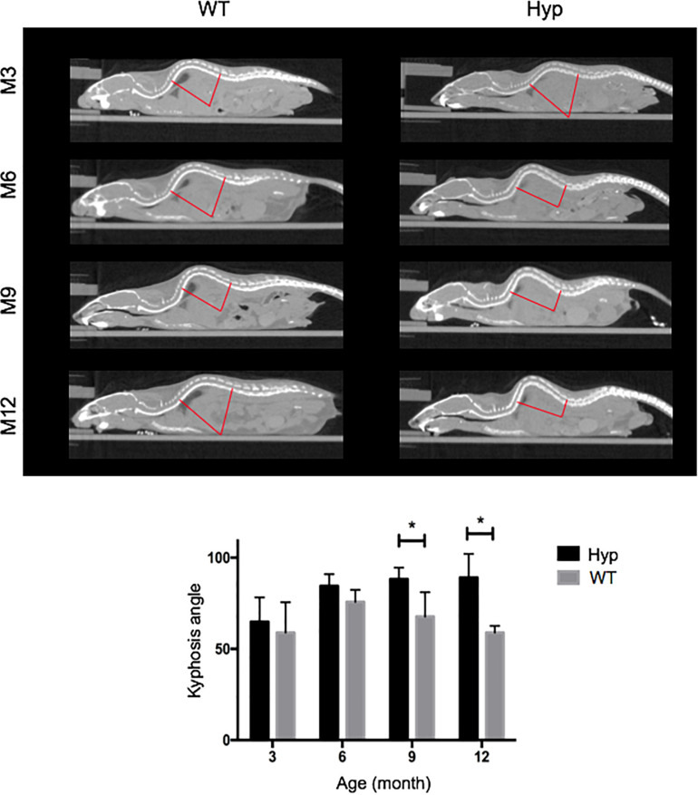
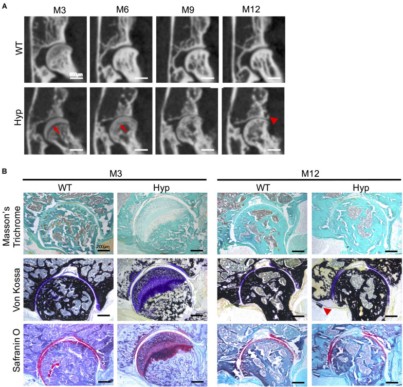
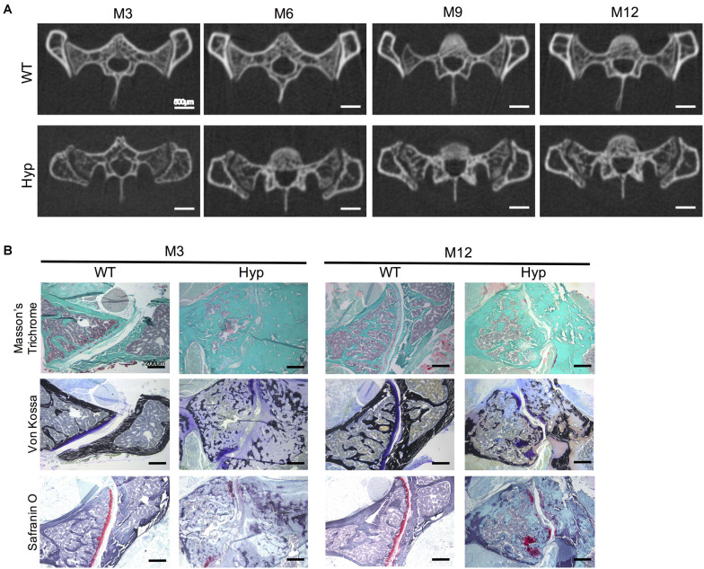
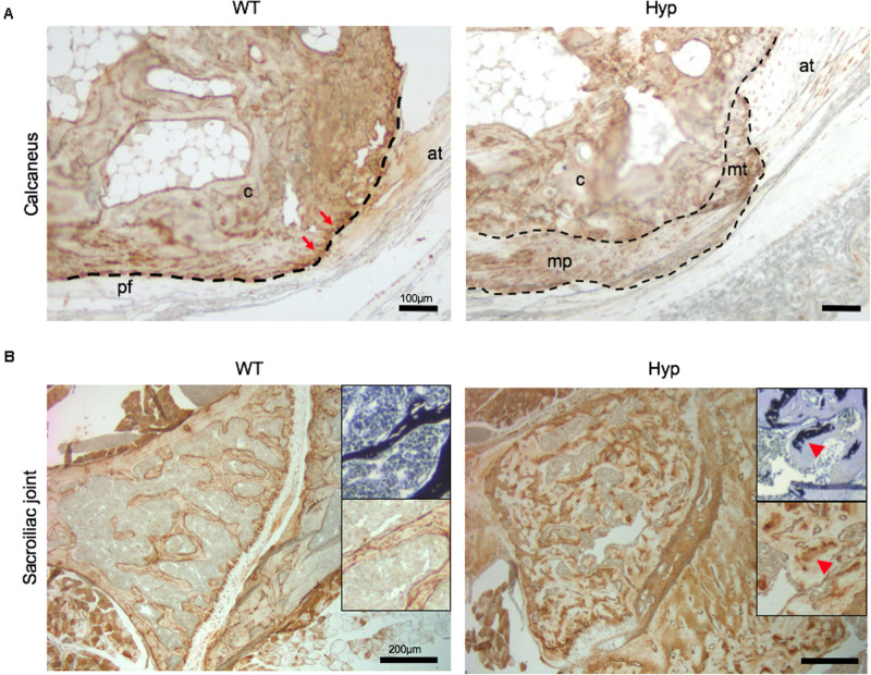
X-linked hypophosphatemia (XLH) is characterized by rickets and osteomalacia, caused by inactivating mutations in the Phosphate-regulating endopeptidase homolog X-linked (PHEX) gene. With aging, adult patients develop paradoxical heterotopic calcifications of tendons and ligaments at their insertion sites (enthesophytes), and joint alterations. Understanding the progression of this structural damage that severely affects patients' quality of life will help to improve the management of XLH. Here, we characterized the occurrence of enthesophytes and joint alterations through a 12 month micro-CT follow-up in the mouse, a murine model of XLH ( = 5 mice per group). Similar to adult patients with XLH, mice developed calcaneal enthesophytes, hip joint alterations, erosions of the sacroiliac joints and periarticular calcifications. These lesions were already present at month 3 and gradually worsened over time. In sharp contrast, no abnormalities were observed in control mice at early time points. Histological analyses confirmed the presence of bone erosions, calcifications and expansion of mineralizing enthesis fibrocartilage in mice and their absence in controls and suggested that new bone formation is driven by altered mechanical strain. Interestingly, despite a strong deformation of the curvature, none of the mice displayed enthesophyte at the spine. Peripheral enthesophytes and joint alterations develop at the early stages of the disease and gradually worsen overtime. Overall, our findings highlight the relevance of this preclinical model to test new therapies aiming to prevent bone and joint complications in XLH.
X连锁低磷血症(XLH)的特征是佝偻病和骨软化症,由X连锁磷酸盐调节内肽酶同源物(PHEX)基因的失活突变引起。随着年龄增长,成年患者在其肌腱和韧带附着部位(附着点骨赘)出现反常的异位钙化以及关节改变。了解这种严重影响患者生活质量的结构损伤的进展情况将有助于改善XLH的治疗管理。在此,我们通过对XLH小鼠模型(每组5只小鼠)进行为期12个月的微型计算机断层扫描(micro-CT)随访,对附着点骨赘和关节改变的发生情况进行了表征。与成年XLH患者相似,XLH小鼠出现了跟骨附着点骨赘、髋关节改变、骶髂关节侵蚀和关节周围钙化。这些病变在3个月时就已出现,并随时间逐渐恶化。与之形成鲜明对比的是,在早期时间点,对照小鼠未观察到任何异常。组织学分析证实,XLH小鼠存在骨侵蚀、钙化以及矿化附着点纤维软骨的扩展,而对照小鼠则没有,这表明新骨形成是由改变的机械应变驱动的。有趣的是,尽管脊柱曲率有强烈变形,但所有XLH小鼠在脊柱处均未出现附着点骨赘。周围附着点骨赘和关节改变在疾病早期出现,并随时间逐渐恶化。总体而言,我们的研究结果凸显了这种临床前模型对于测试旨在预防XLH骨和关节并发症的新疗法的相关性。