Radiology Department, Hospital Naval Almirante Nef, Subida Alesandri S/N., Viña del Mar, Provincia de Valparaíso, Chile.
Radiology Department, Hospital Dr. Eduardo Pereira, Valparaiso, Chile.
Crit Care. 2020 Oct 21;24(1):619. doi: 10.1186/s13054-020-03333-3.
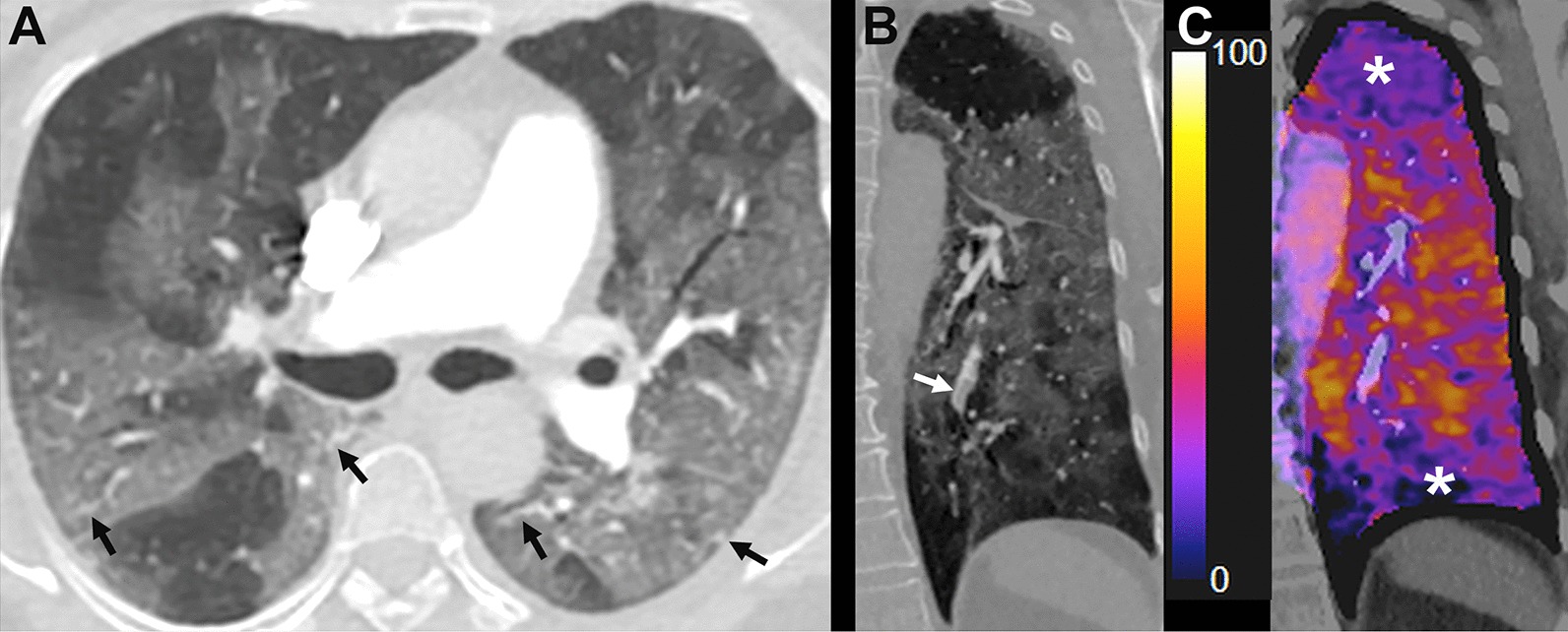
Subtraction CT angiography (sCTA) is a technique used to evaluate pulmonary perfusion based on iodine distribution maps. The aim of this study is to assess lung perfusion changes with sCTA seen in patients with COVID-19 pneumonia and correlate them with clinical outcomes.
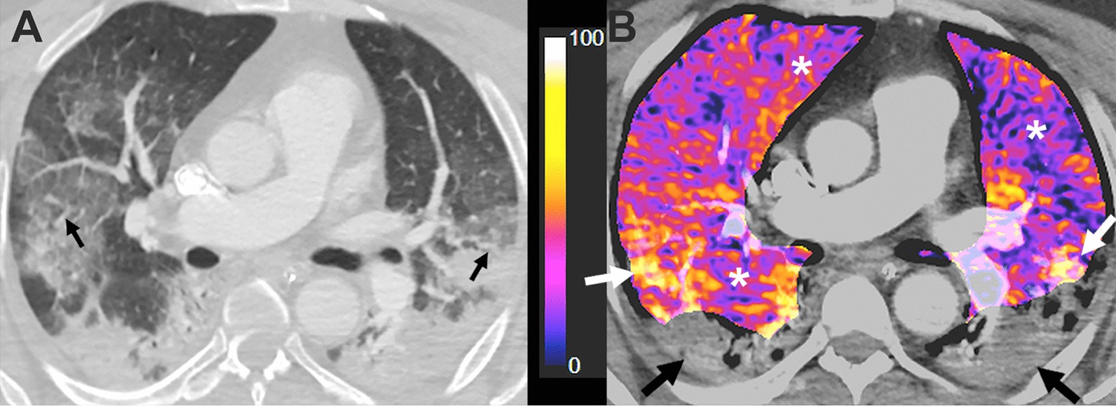
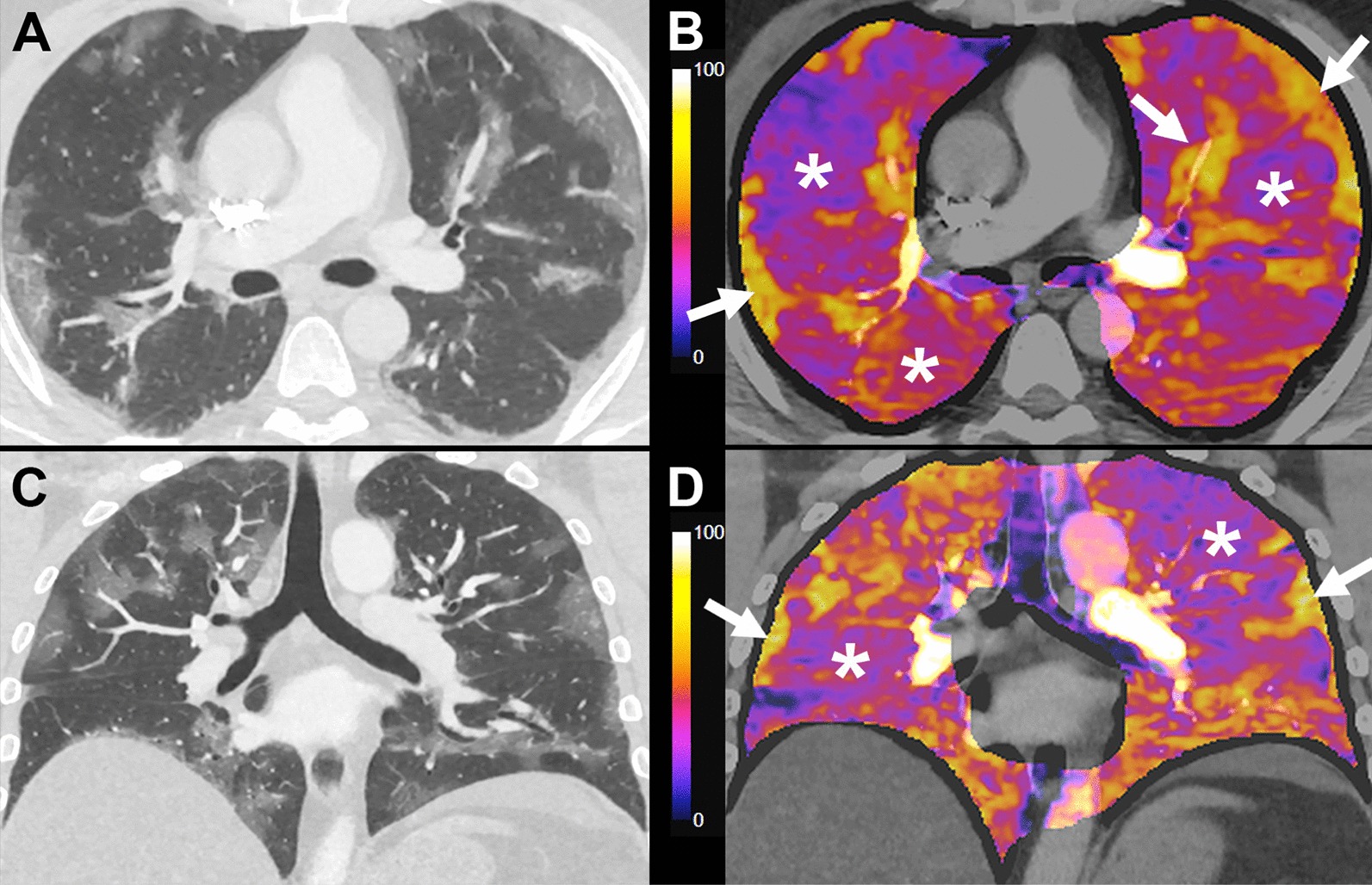
A prospective cohort study was carried out with 45 RT-PCR-confirmed COVID-19 patients that required hospitalization at three different hospitals, between April and May 2020. In all cases, a basic clinical and demographic profile was obtained. Lung perfusion was assessed using sCTA. Evaluated imaging features included: Pattern predominance of injured lung parenchyma in both lungs (ground-glass opacities, consolidation and mixed pattern) and anatomical extension; predominant type of perfusion abnormality (increased perfusion or hypoperfusion), perfusion abnormality distribution (focal or diffuse), extension of perfusion abnormalities (mild, moderate and severe involvement); presence of vascular dilatation and vascular tortuosity. All participants were followed-up until hospital discharge searching for the development of any of the study endpoints. These endpoints included intensive-care unit (ICU) admission, initiation of invasive mechanical ventilation (IMV) and death.
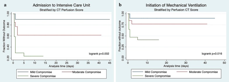
Forty-one patients (55.2 ± 16.5 years, 22 men) with RT-PCR-confirmed SARS-CoV-2 infection and an interpretable iodine map were included. Patients with perfusion anomalies on sCTA in morphologically normal lung parenchyma showed lower Pa/Fi values (294 ± 111.3 vs. 397 ± 37.7, p = 0.035), and higher D-dimer levels (1156 ± 1018 vs. 378 ± 60.2, p < 0.01). The main common patterns seen in lung CT scans were ground-glass opacities, mixed pattern with predominant ground-glass opacities and mixed pattern with predominant consolidation in 56.1%, 24.4% and 19.5% respectively. Perfusion abnormalities were common (36 patients, 87.8%), mainly hypoperfusion in areas of apparently healthy lung. Patients with severe hypoperfusion in areas of apparently healthy lung parenchyma had an increased probability of being admitted to ICU and to initiate IMV (HR of 11.9 (95% CI 1.55-91.9) and HR 7.8 (95% CI 1.05-61.1), respectively).
Perfusion abnormalities evidenced in iodine maps obtained by sCTA are associated with increased admission to ICU and initiation of IMV in COVID-19 patients.
减影 CT 血管造影术(sCTA)是一种基于碘分布图谱评估肺灌注的技术。本研究旨在评估 COVID-19 肺炎患者 sCTA 所见的肺灌注变化,并将其与临床结局相关联。
这是一项前瞻性队列研究,纳入了 2020 年 4 月至 5 月期间在三家不同医院住院的 45 例经 RT-PCR 确诊的 COVID-19 患者。所有患者均获得了基本的临床和人口统计学特征。使用 sCTA 评估肺灌注。评估的影像学特征包括:双肺损伤性肺实质(磨玻璃影、实变和混合影)和解剖学延伸的优势模式;灌注异常的主要类型(灌注增加或灌注减少),灌注异常分布(局灶性或弥漫性),灌注异常的延伸(轻度、中度和重度受累);血管扩张和血管扭曲的存在。所有参与者均随访至出院,以寻找研究终点的任何进展。这些终点包括入住重症监护病房(ICU)、开始有创机械通气(IMV)和死亡。
共纳入 41 例(55.2±16.5 岁,22 名男性)经 RT-PCR 确诊 SARS-CoV-2 感染且碘图可解读的患者。sCTA 上在形态正常的肺实质中出现灌注异常的患者,其 Pa/Fi 值较低(294±111.3 与 397±37.7,p=0.035),D-二聚体水平较高(1156±1018 与 378±60.2,p<0.01)。肺部 CT 扫描的主要常见模式为磨玻璃影、以磨玻璃影为主的混合影和以实变为主的混合影,分别占 56.1%、24.4%和 19.5%。灌注异常很常见(36 例,87.8%),主要表现为看似健康的肺区灌注减少。在看似健康的肺实质区域出现严重灌注减少的患者,入住 ICU 和开始 IMV 的概率增加(HR 为 11.9(95%CI 1.55-91.9)和 HR 为 7.8(95%CI 1.05-61.1))。
sCTA 碘图所示的灌注异常与 COVID-19 患者入住 ICU 和开始 IMV 的概率增加相关。