School of Life Sciences, Tsinghua-Peking Joint Center for Life Sciences, Beijing Advanced Innovation Center for Structural Biology, Beijing Frontier Research Center for Biological Structure, Tsinghua University, 100084 Beijing, China.
School of Life Sciences, Tsinghua-Peking Joint Center for Life Sciences, Beijing Advanced Innovation Center for Structural Biology, Beijing Frontier Research Center for Biological Structure, Tsinghua University, 100084 Beijing, China
Proc Natl Acad Sci U S A. 2020 Nov 3;117(44):27124-27131. doi: 10.1073/pnas.2008447117. Epub 2020 Oct 21.
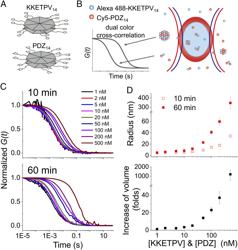
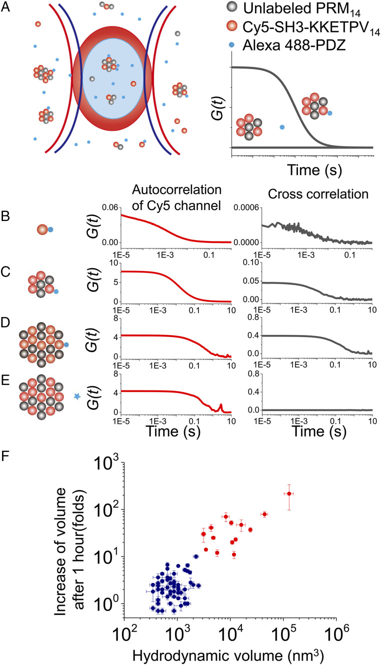
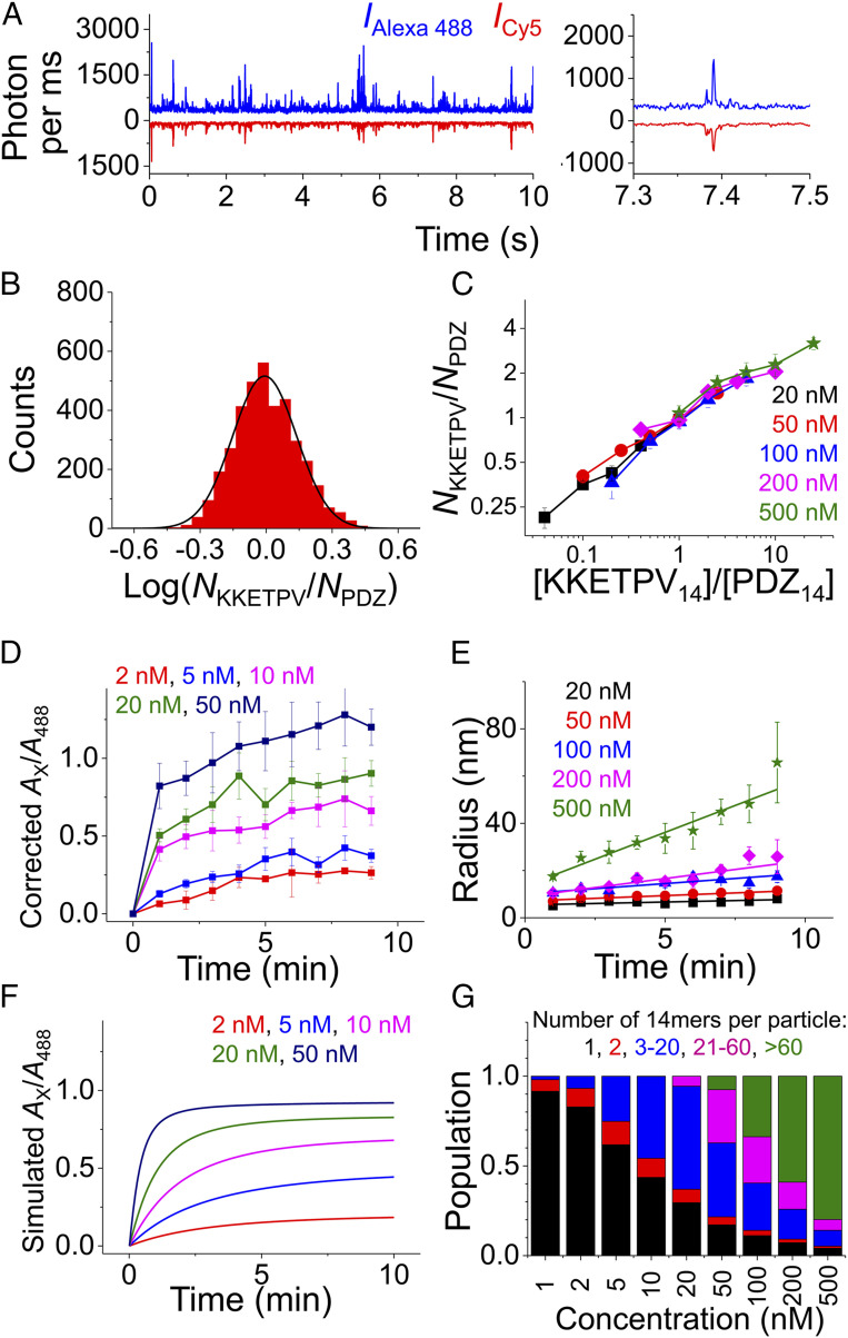
Liquid-liquid phase separation, driven by multivalent macromolecular interactions, causes formation of membraneless compartments, which are biomolecular condensates containing concentrated macromolecules. These condensates are essential in diverse cellular processes. Formation and dynamics of micrometer-scale phase-separated condensates are examined routinely. However, limited by commonly used methods which cannot capture small-sized free-diffusing condensates, the transition process from miscible individual molecules to micrometer-scale condensates is mostly unknown. Herein, with a dual-color fluorescence cross-correlation spectroscopy (dcFCCS) method, we captured formation of nanoscale condensates beyond the detection limit of conventional fluorescence microscopy. In addition, dcFCCS is able to quantify size and growth rate of condensates as well as molecular stoichiometry and binding affinity of client molecules within condensates. The critical concentration to form nanoscale condensates, identified by our experimental measurements and Monte Carlo simulations, is at least several fold lower than the detection limit of conventional fluorescence microscopy. Our results emphasize that, in addition to micrometer-scale condensates, nanoscale condensates are likely to play important roles in various cellular processes and dcFCCS is a simple and powerful quantitative tool to examine them in detail.
液-液相分离受多价大分子相互作用驱动,导致无膜隔室的形成,这些无膜隔室是含有浓缩大分子的生物分子凝聚体。这些凝聚体在各种细胞过程中是必不可少的。微米尺度的相分离凝聚体的形成和动力学通常是可以检测到的。然而,由于常用的方法不能捕获小尺寸的自由扩散凝聚体,从可混溶的单个分子到微米尺度的凝聚体的转变过程在很大程度上是未知的。在此,我们采用双色荧光相关光谱(dcFCCS)方法,捕获了常规荧光显微镜无法检测到的纳米级凝聚体的形成。此外,dcFCCS 能够定量地确定凝聚体的大小和生长速率,以及凝聚体中客户分子的分子化学计量和结合亲和力。通过实验测量和蒙特卡罗模拟确定的形成纳米级凝聚体的临界浓度至少比常规荧光显微镜的检测极限低几个数量级。我们的研究结果强调,除了微米尺度的凝聚体之外,纳米尺度的凝聚体可能在各种细胞过程中发挥重要作用,dcFCCS 是一种简单而强大的定量工具,可以详细地检测它们。