Department of Gastroenterology, Japanese Red Cross Nagoya Daini Hospital, Japan.
Division of Gastroenterology and Hepatology, University of Iowa, USA.
Intern Med. 2021 Mar 15;60(6):859-866. doi: 10.2169/internalmedicine.5920-20. Epub 2020 Oct 21.
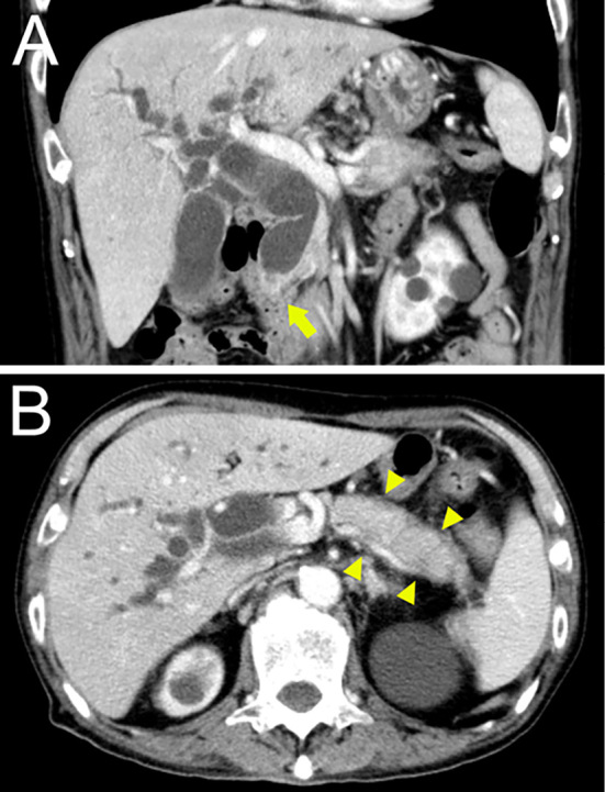
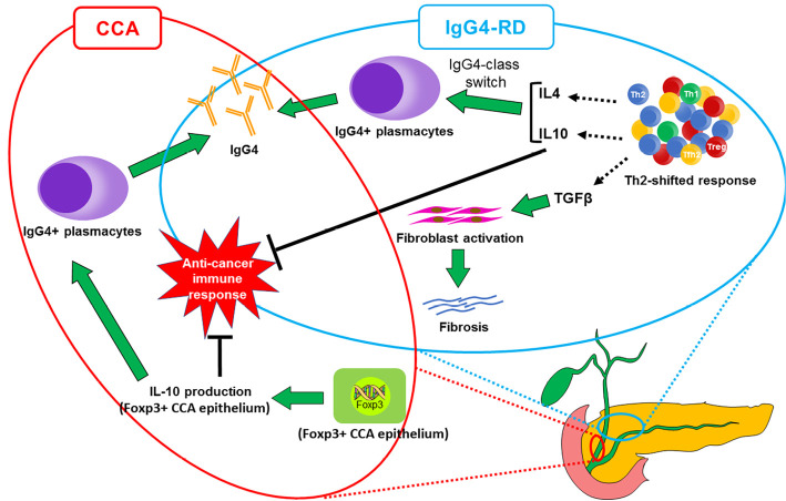
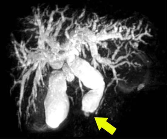
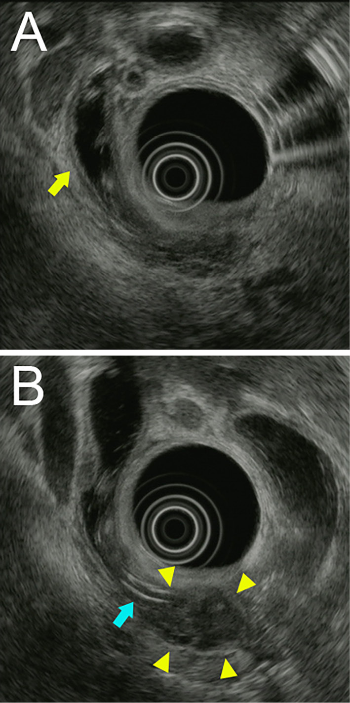
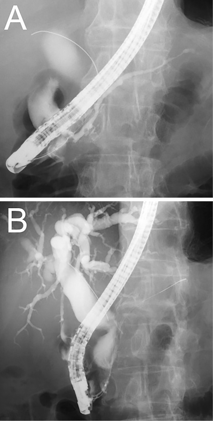
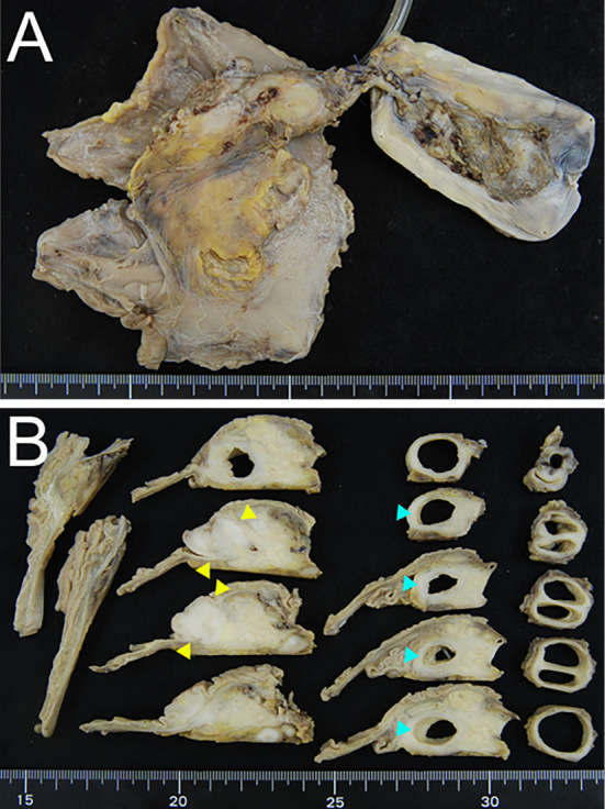
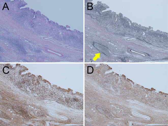
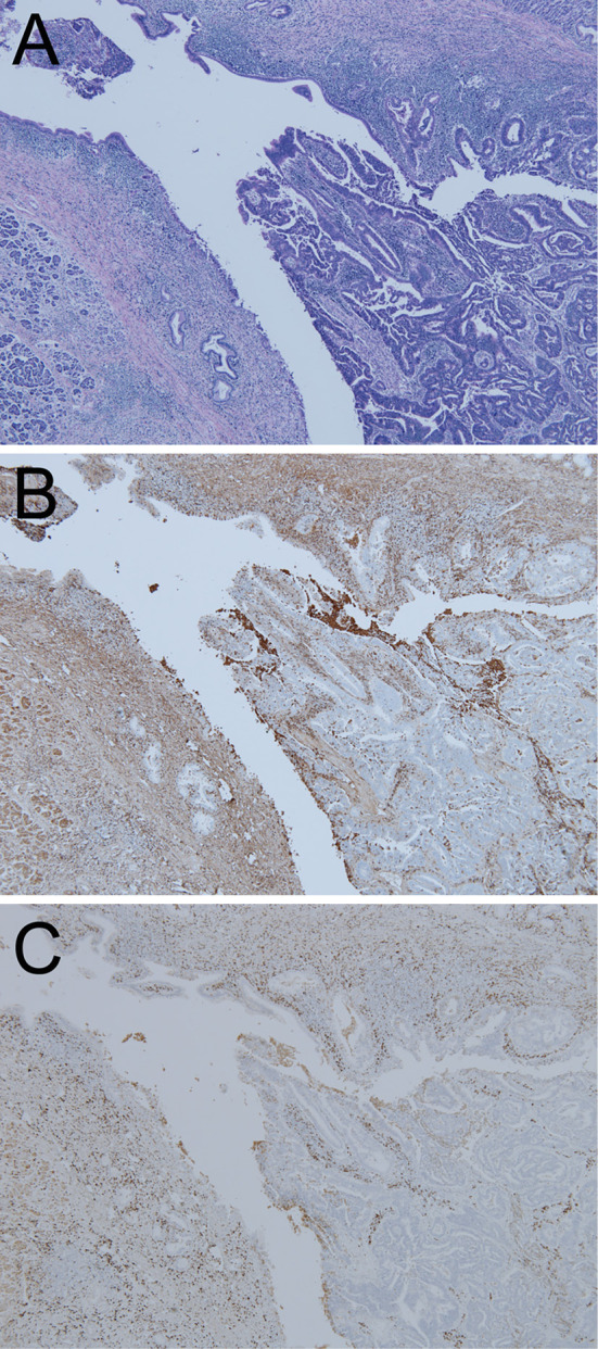
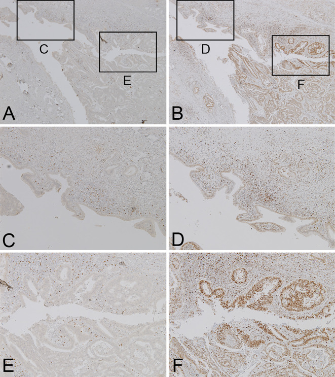
An 80-year-old man was admitted due to biliary stricture with autoimmune pancreatitis. Although radiographical examinations suggested Immunoglobulin G4-related sclerosing cholangitis (IgG4-SC), punched biopsies from the bile duct revealed adenocarcinoma. In the resected specimen, abundant N-terminus of Forkhead box P3 (Foxp3)-positive cells were localized in cholangiocarcinoma (CCA) tissue, while IgG4-positive cells were spread around the entire bile duct. Therefore, the case was diagnosed with IgG4-SC accompanied by CCA, not sporadic CCA. We herein report an informative case wherein IgG4-positive cells were abundant in CCA tissue and Foxp3 immunohistochemical staining allowed us to determine that this case had two entities.
一位 80 岁男性因胆胰管狭窄伴自身免疫性胰腺炎入院。尽管影像学检查提示 IgG4 相关硬化性胆管炎(IgG4-SC),但胆管穿刺活检显示为腺癌。在切除标本中,大量 Foxp3 阳性细胞位于胆癌(CCA)组织中,而 IgG4 阳性细胞则分布在整个胆管周围。因此,该病例被诊断为 IgG4-SC 伴 CCA,而非散发性 CCA。我们在此报告一例信息丰富的病例,其中 IgG4 阳性细胞在 CCA 组织中丰富,Foxp3 免疫组化染色可确定该病例存在两种实体。