• 文献检索
  • 文档翻译
  • 深度研究
  • 学术资讯
  • Suppr Zotero 插件Zotero 插件
  • 邀请有礼
  • 套餐&价格
  • 历史记录
应用&插件
Suppr Zotero 插件Zotero 插件浏览器插件Mac 客户端Windows 客户端微信小程序
定价
高级版会员购买积分包购买API积分包
服务
文献检索文档翻译深度研究API 文档MCP 服务
关于我们
关于 Suppr公司介绍联系我们用户协议隐私条款
关注我们

Suppr 超能文献

核心技术专利:CN118964589B侵权必究
粤ICP备2023148730 号-1Suppr @ 2026

文献检索

告别复杂PubMed语法,用中文像聊天一样搜索,搜遍4000万医学文献。AI智能推荐,让科研检索更轻松。

立即免费搜索

文件翻译

保留排版,准确专业,支持PDF/Word/PPT等文件格式,支持 12+语言互译。

免费翻译文档

深度研究

AI帮你快速写综述,25分钟生成高质量综述,智能提取关键信息,辅助科研写作。

立即免费体验

评估 OCT-A 设备和扫描范围对视网膜血管参数的影响。

Assessing the Influence of OCT-A Device and Scan Size on Retinal Vascular Metrics.

机构信息

School of Medicine, Medical College of Wisconsin, Milwaukee, WI, USA.

Department of Ophthalmology and Visual Sciences, Medical College of Wisconsin, Milwaukee, WI, USA.

出版信息

Transl Vis Sci Technol. 2020 Oct 7;9(11):7. doi: 10.1167/tvst.9.11.7. eCollection 2020 Oct.

DOI:10.1167/tvst.9.11.7
PMID:33101784
原文链接:https://pmc.ncbi.nlm.nih.gov/articles/PMC7545061/
Abstract

PURPOSE

The purpose of this study was to investigate the effect of device and scan size on quantitative optical coherence tomography angiography (OCT-A) metrics.

METHODS

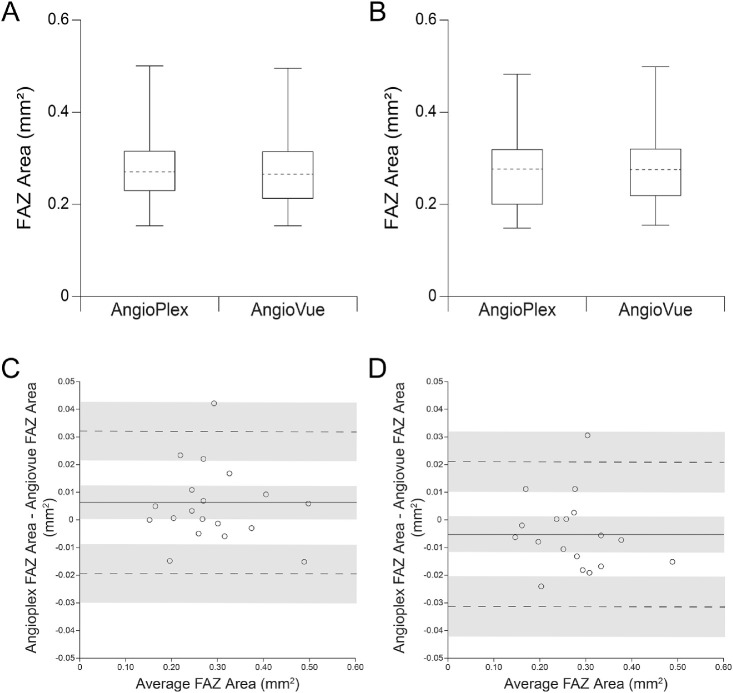
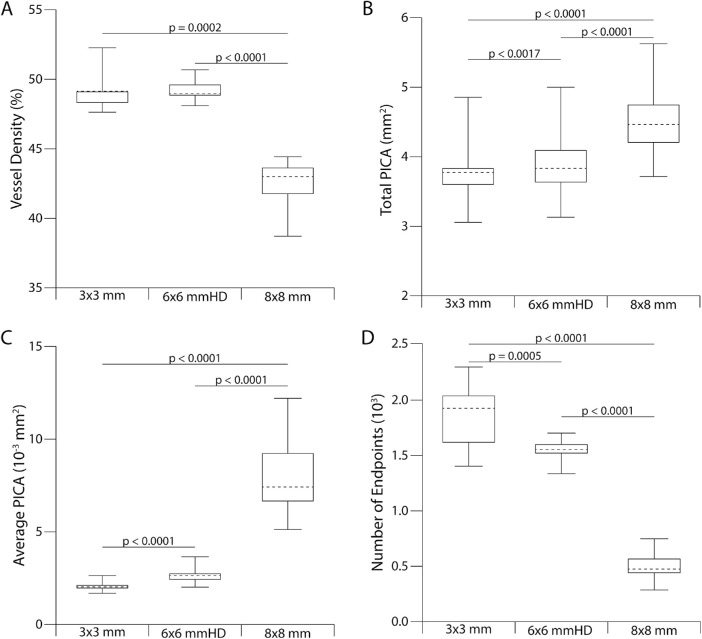
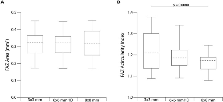
The 3 × 3 mm scans from Optovue AngioVue and Zeiss AngioPlex systems were included for 18 eyes of 18 subjects without ocular pathology. The foveal avascular zone (FAZ) was segmented manually by two observers, from which estimates of FAZ area (using both the nominal image scale and the axial length corrected image scale) and acircularity were derived. Three scan sizes (3 mm, 6 mm HD, and 8 mm) from the AngioVue system were included for 15 eyes of 15 subjects without ocular pathology. For each subject, larger image sizes were resized to the same resolution as 3 × 3 mm scans, aligned, then cropped to a common area. FAZ area, FAZ acircularity, average and total parafoveal intercapillary area, vessel density, and vessel end points were computed.

RESULTS

Between the devices used here, there were no significant differences in FAZ acircularity ( = 0.88) or FAZ area using scaled ( = 0.11) or unscaled images ( = 0.069). Although there was no significant difference in FAZ area across scan sizes ( = 0.30), vessel morphometry metrics were all significantly influenced by scan size.

CONCLUSIONS

The scan devices and sizes used here do not affect FAZ area measures derived from manual segmentations. In contrast, vessel morphometry metrics are affected by scan size. As individual differences in axial length induce differences in absolute scan size, extreme care should be taken when interpreting metrics of vessel morphometry, both between and within OCT-A devices.

TRANSLATIONAL RELEVANCE

A better characterization of the confounds surrounding OCT-A retinal vasculature metrics can lead to improved application of these metrics as biomarkers for retinal and systemic diseases.

摘要

目的

本研究旨在探讨设备和扫描尺寸对定量光相干断层扫描血管造影(OCT-A)指标的影响。

方法

纳入 18 名受试者的 18 只眼,无眼部病变,获取 Optovue AngioVue 和 Zeiss AngioPlex 系统的 3×3mm 扫描。由两名观察者手动分割黄斑中心无血管区(FAZ),从 FAZ 面积(使用标称图像比例和轴向长度校正的图像比例)和非圆度的估计值。纳入 15 名无眼部病变受试者的 15 只眼,获取 AngioVue 系统的三种扫描尺寸(3mm、6mmHD 和 8mm)。对于每个受试者,较大的图像尺寸均按与 3×3mm 扫描相同的分辨率进行调整,然后对齐并裁剪到公共区域。计算 FAZ 面积、FAZ 非圆度、平均和总旁中心毛细血管区、血管密度和血管终点。

结果

在此使用的设备之间,FAZ 非圆度( = 0.88)或使用缩放( = 0.11)或未缩放图像( = 0.069)的 FAZ 面积无显著差异。尽管扫描尺寸之间 FAZ 面积无显著差异( = 0.30),但血管形态计量学指标均受扫描尺寸影响。

结论

在此使用的扫描设备和尺寸不会影响手动分割得出的 FAZ 面积测量值。相反,血管形态计量学指标受扫描尺寸影响。由于眼轴长度的个体差异导致绝对扫描尺寸的差异,因此在解释 OCT-A 血管形态计量学的指标时,无论是在 OCT-A 设备之间还是内部,都应格外小心。

翻译

于婷

https://cdn.ncbi.nlm.nih.gov/pmc/blobs/17f5/7545061/76bbdb87f23c/tvst-9-11-7-f004.jpg
https://cdn.ncbi.nlm.nih.gov/pmc/blobs/17f5/7545061/07e0dc223672/tvst-9-11-7-f001.jpg
https://cdn.ncbi.nlm.nih.gov/pmc/blobs/17f5/7545061/6e5ea0cd531f/tvst-9-11-7-f002.jpg
https://cdn.ncbi.nlm.nih.gov/pmc/blobs/17f5/7545061/c4bb7404f769/tvst-9-11-7-f003.jpg
https://cdn.ncbi.nlm.nih.gov/pmc/blobs/17f5/7545061/76bbdb87f23c/tvst-9-11-7-f004.jpg

相似文献

1
Assessing the Influence of OCT-A Device and Scan Size on Retinal Vascular Metrics.评估 OCT-A 设备和扫描范围对视网膜血管参数的影响。
Transl Vis Sci Technol. 2020 Oct 7;9(11):7. doi: 10.1167/tvst.9.11.7. eCollection 2020 Oct.
2
The Impact of Axial Eye Growth on Foveal Avascular Zone Measurements in Children.轴向眼球生长对儿童中心凹无血管区测量的影响。
Optom Vis Sci. 2022 Feb 1;99(2):127-136. doi: 10.1097/OPX.0000000000001854.
3
Normative Data Assessment of Peripapillary and Macular Vessel Density and Foveal Avascular Zone Metrics Using Optical Coherence Tomography Angiography in Children.使用光学相干断层血管造影术评估儿童视乳头周围和黄斑血管密度及中心无血管区参数的正常值。
J Pediatr Ophthalmol Strabismus. 2020 Nov 1;57(6):388-398. doi: 10.3928/01913913-20200903-01.
4
[Comparison Between Automated and Manual Measurement of Foveal Avascular Zone Retinopathy in Optical Coherence Tomography].光学相干断层扫描中黄斑无血管区视网膜病变的自动测量与手动测量的比较
Klin Monbl Augenheilkd. 2019 Dec;236(12):1445-1450. doi: 10.1055/a-1002-0045. Epub 2019 Oct 31.
5
Comparative Evaluation of Foveal Avascular Zone on Two Optical Coherence Tomography Angiography Devices.两种光学相干断层扫描血管造影设备对黄斑无血管区的比较评估
Optom Vis Sci. 2018 Jul;95(7):602-607. doi: 10.1097/OPX.0000000000001238.
6
[Correlation of capillary plexus with visual acuity in idiopathic macular epiretinal membrane eyes using optical coherence tomography angiography].[利用光学相干断层扫描血管造影术研究特发性黄斑视网膜前膜眼中毛细血管丛与视力的相关性]
Zhonghua Yan Ke Za Zhi. 2019 Oct 11;55(10):757-762. doi: 10.3760/cma.j.issn.0412-4081.2019.10.006.
7
Optical coherence tomography angiography of superficial retinal vessel density and foveal avascular zone in myopic children.近视儿童的浅层视网膜血管密度和中心无血管区的光相干断层扫描血管造影。
PLoS One. 2019 Jul 18;14(7):e0219785. doi: 10.1371/journal.pone.0219785. eCollection 2019.
8
Assessing the Sensitivity of OCT-A Retinal Vasculature Metrics.评估 OCT-A 视网膜血管测量指标的敏感性。
Transl Vis Sci Technol. 2023 Aug 1;12(8):2. doi: 10.1167/tvst.12.8.2.
9
VARIABILITY IN FOVEAL AVASCULAR ZONE AND CAPILLARY DENSITY USING OPTICAL COHERENCE TOMOGRAPHY ANGIOGRAPHY MACHINES IN HEALTHY EYES.使用光学相干断层扫描血管造影机检测健康眼睛的黄斑中心凹血管区和毛细血管密度的变异性
Retina. 2017 Nov;37(11):2102-2111. doi: 10.1097/IAE.0000000000001458.
10
OPTICAL COHERENCE TOMOGRAPHY ANGIOGRAPHY OF THE FOVEA IN CHILDREN BORN PRETERM.早产儿黄斑区的光学相干断层扫描血管造影
Retina. 2017 Dec;37(12):2289-2294. doi: 10.1097/IAE.0000000000001471.

引用本文的文献

1
Unlocking the Potential of Vessel Density and the Foveal Avascular Zone in Optical Coherence Tomography Angiography as Biomarkers in Alzheimer's Disease.揭示光学相干断层扫描血管造影中血管密度和黄斑无血管区作为阿尔茨海默病生物标志物的潜力。
Healthcare (Basel). 2024 Aug 9;12(16):1589. doi: 10.3390/healthcare12161589.
2
In Vivo Assessment of Retinal Phenotypes in Axenfeld-Rieger Syndrome.体内评估 Axenfeld-Rieger 综合征的视网膜表型。
Invest Ophthalmol Vis Sci. 2024 Apr 1;65(4):20. doi: 10.1167/iovs.65.4.20.
3
The Shape of the Foveal Avascular Zone: When a Circle Isn't Round.

本文引用的文献

1
Visual Acuity and Foveal Structure in Eyes with Fragmented Foveal Avascular Zones.伴有部分性黄斑区无血管区的眼的视力和中心凹结构。
Ophthalmol Retina. 2020 May;4(5):535-544. doi: 10.1016/j.oret.2019.11.014. Epub 2019 Nov 22.
2
OCTA: A Practical Method of Image Averaging Using Adobe Photoshop Software.光学相干断层扫描血管造影术:一种使用Adobe Photoshop软件进行图像平均处理的实用方法。
Ophthalmic Surg Lasers Imaging Retina. 2019 Dec 1;50(12):802-807. doi: 10.3928/23258160-20191119-09.
3
Comparison of the Repeatability of Macular Vascular Density Measurements Using Four Optical Coherence Tomography Angiography Systems.
黄斑中心凹无血管区的形态:圆非其圆。
Transl Vis Sci Technol. 2023 Jun 1;12(6):26. doi: 10.1167/tvst.12.6.26.
4
Retinal imaging biomarkers of Alzheimer's disease: A systematic review and meta-analysis of studies using brain amyloid beta status for case definition.阿尔茨海默病的视网膜成像生物标志物:一项使用脑淀粉样蛋白β状态进行病例定义的研究的系统评价和荟萃分析。
Alzheimers Dement (Amst). 2023 May 25;15(2):e12421. doi: 10.1002/dad2.12421. eCollection 2023 Apr-Jun.
5
Racial differences in quantitative optical coherence tomography angiography findings between older non-diabetics with co-morbidities.老年人合并症中非糖尿病患者定量光学相干断层扫描血管造影结果的种族差异。
PLoS One. 2023 May 5;18(5):e0285360. doi: 10.1371/journal.pone.0285360. eCollection 2023.
6
Diurnal changes in choroidal optical coherence tomography angiography indices over 24 hours in healthy young adults.健康年轻成年人 24 小时内脉络膜光学相干断层扫描血管造影指数的昼夜变化。
Sci Rep. 2023 Mar 2;13(1):3551. doi: 10.1038/s41598-023-30433-1.
7
Assessing Foveal Structure in Individuals with R402Q and S192Y Hypomorphic Alleles.评估具有R402Q和S192Y次等位基因个体的中央凹结构。
Ophthalmol Sci. 2021 Nov 17;1(4):100077. doi: 10.1016/j.xops.2021.100077. eCollection 2021 Dec.
8
Changes in Retinal Optical Coherence Tomography Angiography Indexes Over 24 Hours.视网膜光学相干断层扫描血管造影指数 24 小时内的变化。
Invest Ophthalmol Vis Sci. 2022 Mar 2;63(3):25. doi: 10.1167/iovs.63.3.25.
9
OCTAVA: An open-source toolbox for quantitative analysis of optical coherence tomography angiography images.OCTAVA:用于光学相干断层扫描血管造影图像定量分析的开源工具箱。
PLoS One. 2021 Dec 9;16(12):e0261052. doi: 10.1371/journal.pone.0261052. eCollection 2021.
10
Early-stage macular telangiectasia type 2 vascular abnormalities are associated with interdigitation zone disruption.早期 2 型黄斑毛细血管扩张症的血管异常与交织区破坏有关。
PLoS One. 2021 Nov 12;16(11):e0259811. doi: 10.1371/journal.pone.0259811. eCollection 2021.
使用四种光学相干断层扫描血管造影系统对黄斑血管密度测量的可重复性比较。
J Ophthalmol. 2019 Nov 23;2019:4372580. doi: 10.1155/2019/4372580. eCollection 2019.
4
Assessing the Use of Incorrectly Scaled Optical Coherence Tomography Angiography Images in Peer-Reviewed Studies: A Systematic Review.评估同行评审研究中使用错误缩放光学相干断层扫描血管造影图像的情况:系统评价。
JAMA Ophthalmol. 2020 Jan 1;138(1):86-94. doi: 10.1001/jamaophthalmol.2019.4821.
5
Microaneurysm Imaging Using Multiple En Face OCT Angiography Image Averaging: Morphology and Visualization.多层面 OCT 血管成像平均法检测微动脉瘤:形态与可视化。
Ophthalmol Retina. 2020 Feb;4(2):175-186. doi: 10.1016/j.oret.2019.09.010. Epub 2019 Sep 27.
6
Quantitative Comparison Of Microvascular Metrics On Three Optical Coherence Tomography Angiography Devices In Chorioretinal Disease.三种光学相干断层扫描血管造影设备在脉络膜视网膜疾病中微血管指标的定量比较
Clin Ophthalmol. 2019 Oct 22;13:2063-2069. doi: 10.2147/OPTH.S215322. eCollection 2019.
7
Age and gender influence on foveal avascular zone in healthy eyes.年龄和性别对健康眼中中心凹无血管区的影响。
Exp Eye Res. 2019 Dec;189:107856. doi: 10.1016/j.exer.2019.107856. Epub 2019 Oct 22.
8
Impact of integrated multiple image averaging on OCT angiography image quality and quantitative parameters.整合多幅图像平均对 OCT 血管造影图像质量和定量参数的影响。
Graefes Arch Clin Exp Ophthalmol. 2019 Dec;257(12):2623-2629. doi: 10.1007/s00417-019-04492-y. Epub 2019 Oct 19.
9
Quantification of Choriocapillaris with Optical Coherence Tomography Angiography: A Comparison Study.脉络膜毛细血管的光学相干断层扫描血管造影定量分析:一项对比研究。
Am J Ophthalmol. 2019 Dec;208:111-123. doi: 10.1016/j.ajo.2019.07.003. Epub 2019 Jul 16.
10
OCT Angiography in Young Children with a History of Retinopathy of Prematurity.患有早产儿视网膜病变病史的幼儿的光学相干断层扫描血管造影
Ophthalmol Retina. 2018 Sep;2(9):972-978. doi: 10.1016/j.oret.2018.02.004. Epub 2018 Jul 20.