• 文献检索
  • 文档翻译
  • 深度研究
  • 学术资讯
  • Suppr Zotero 插件Zotero 插件
  • 邀请有礼
  • 套餐&价格
  • 历史记录
应用&插件
Suppr Zotero 插件Zotero 插件浏览器插件Mac 客户端Windows 客户端微信小程序
定价
高级版会员购买积分包购买API积分包
服务
文献检索文档翻译深度研究API 文档MCP 服务
关于我们
关于 Suppr公司介绍联系我们用户协议隐私条款
关注我们

Suppr 超能文献

核心技术专利:CN118964589B侵权必究
粤ICP备2023148730 号-1Suppr @ 2026

文献检索

告别复杂PubMed语法,用中文像聊天一样搜索,搜遍4000万医学文献。AI智能推荐,让科研检索更轻松。

立即免费搜索

文件翻译

保留排版,准确专业,支持PDF/Word/PPT等文件格式,支持 12+语言互译。

免费翻译文档

深度研究

AI帮你快速写综述,25分钟生成高质量综述,智能提取关键信息,辅助科研写作。

立即免费体验

基于 [Formula: see text]-制备的全脑 pCASL 的定量横向弛豫时间图的可靠性。

Reliability of quantitative transverse relaxation time mapping with [Formula: see text]-prepared whole brain pCASL.

机构信息

Department of Epileptology, University of Bonn Medical Center, Bonn, Germany.

German Center for Neurodegenerative Diseases (DZNE), Bonn, Germany.

出版信息

Sci Rep. 2020 Oct 27;10(1):18299. doi: 10.1038/s41598-020-74680-y.

DOI:10.1038/s41598-020-74680-y
PMID:33110203
原文链接:https://pmc.ncbi.nlm.nih.gov/articles/PMC7592060/
Abstract

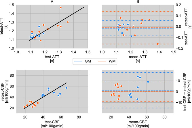
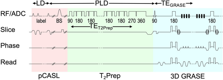
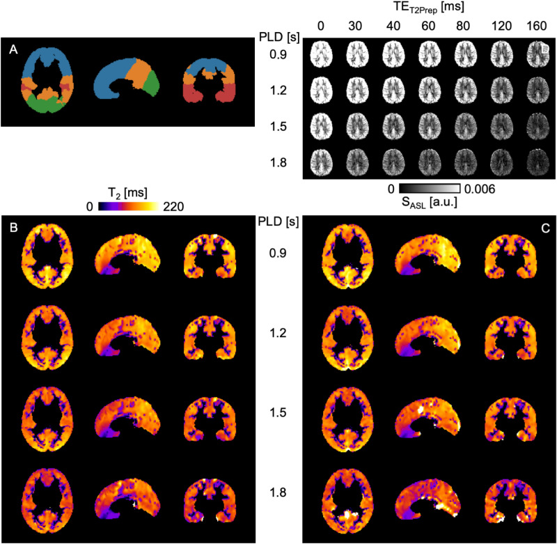
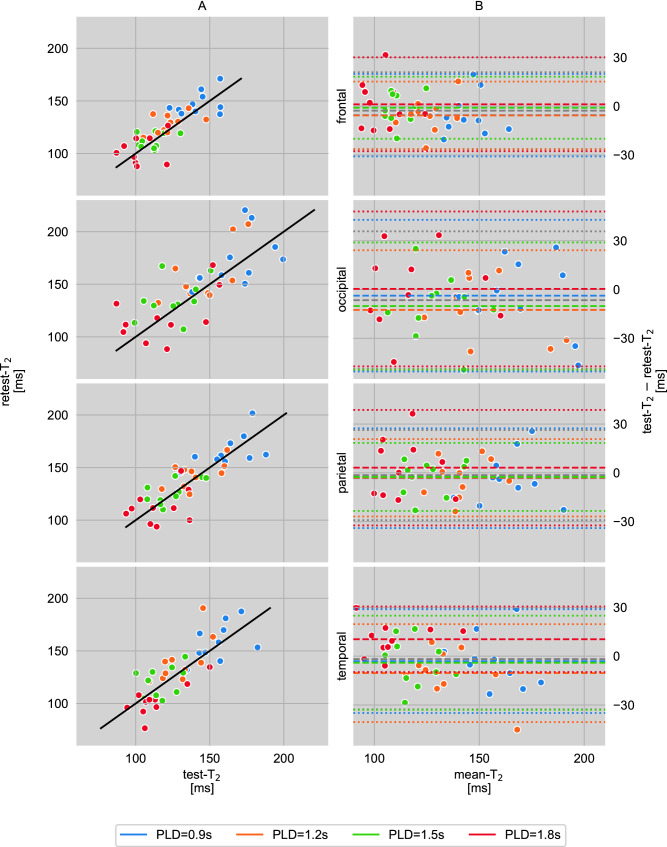
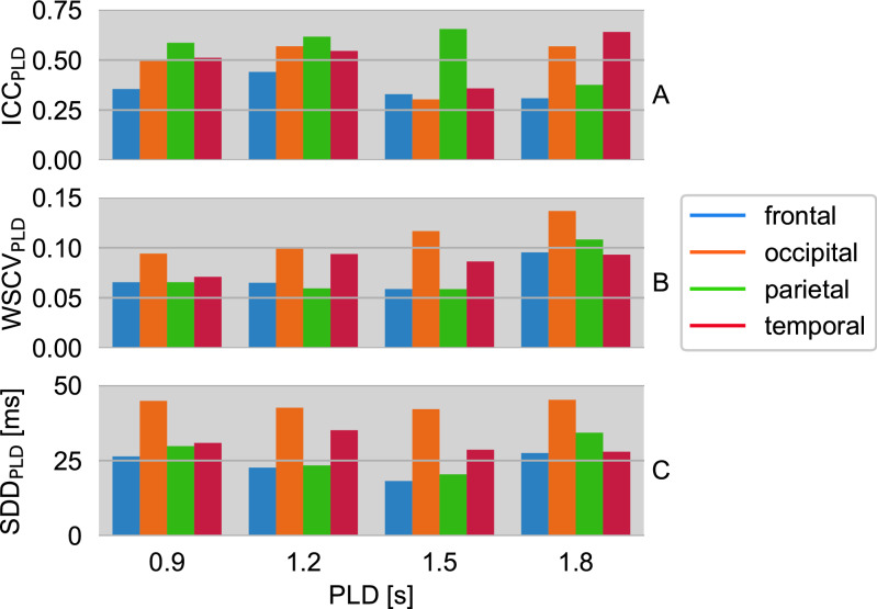
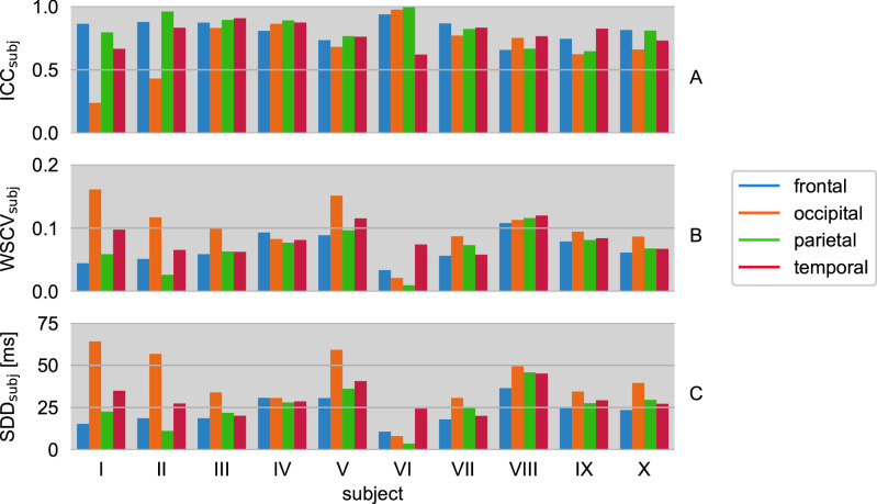
Arterial spin labeling (ASL) is increasingly applied for cerebral blood flow mapping, but [Formula: see text] relaxation of the ASL signal magnetization is often ignored, although it may be clinically relevant. To investigate the extent, to which quantitative [Formula: see text] values in gray matter (GM) obtained by pseudocontinuous ASL (pCASL) perfusion MRI can be reproduced, are reliable and a potential neuroscientific biomarker, a prospective study was performed with ten healthy volunteers (5F,28 ± 3y) at a 3 T scanner. A [Formula: see text]-prepared pCASL sequence enabled the measurement of quantitative [Formula: see text] and perfusion maps. [Formula: see text] times were modeled per voxel and analyzed within four GM-regions-of-interest (ROI). The intraclass correlation coefficients (ICCs) of the quantified ASL-[Formula: see text] varied across brain regions. When averaged across subjects and postlabeling delays (PLDs), the ICCs ranged from reasonable values in parietal regions (ICC = 0.56) to smaller values in frontal regions (ICC = 0.36). Corresponding subject-averaged within-subject coefficients of variation (WSCVs) showed good test-retest measurement precision ([Formula: see text] for all PLDs), but more pronounced inter-subject variance. Reliability and precision of quantified ASL-[Formula: see text] were region-, PLD- and subject-specific, showing fair to robust results in occipital, parietal and temporal ROIs. The results give rise to consider the method for future cerebral studies, where variable perfusion or altered [Formula: see text] times are suspected.

摘要

动脉自旋标记 (ASL) 越来越多地应用于脑血流mapping,但 ASL 信号磁化率的[Formula: see text]弛豫通常被忽略,尽管它可能具有临床相关性。为了研究通过伪连续 ASL (pCASL) 灌注 MRI 获得的灰质 (GM) 中的定量[Formula: see text]值在多大程度上可以重现、可靠且是潜在的神经科学生物标志物,我们在 3T 扫描仪上对 10 名健康志愿者(5 名女性,28±3 岁)进行了前瞻性研究。[Formula: see text] 准备的 pCASL 序列可用于测量定量[Formula: see text]和灌注图。对每个体素进行[Formula: see text]时间建模,并在四个 GM 感兴趣区 (ROI) 内进行分析。定量 ASL-[Formula: see text]的组内相关系数 (ICC) 在脑区之间有所不同。当平均跨受试者和标记后延迟 (PLD) 时,ICC 范围从顶叶区域的合理值(ICC=0.56)到额叶区域的较小值(ICC=0.36)。相应的受试者平均个体内变异系数 (WSCV) 显示出良好的测试-重测测量精度 ([Formula: see text]对于所有 PLD),但个体间变异性更大。定量 ASL-[Formula: see text]的可靠性和精度具有区域、PLD 和受试者特异性,在枕叶、顶叶和颞叶 ROI 中表现出良好到稳健的结果。这些结果表明,在怀疑存在可变灌注或改变的[Formula: see text]时间的情况下,该方法可用于未来的大脑研究。

https://cdn.ncbi.nlm.nih.gov/pmc/blobs/a032/7592060/bfbaa5f3fbcb/41598_2020_74680_Fig7_HTML.jpg
https://cdn.ncbi.nlm.nih.gov/pmc/blobs/a032/7592060/732dda695206/41598_2020_74680_Fig1_HTML.jpg
https://cdn.ncbi.nlm.nih.gov/pmc/blobs/a032/7592060/68a3d8ee58e0/41598_2020_74680_Fig2_HTML.jpg
https://cdn.ncbi.nlm.nih.gov/pmc/blobs/a032/7592060/e159b6ebd285/41598_2020_74680_Fig3_HTML.jpg
https://cdn.ncbi.nlm.nih.gov/pmc/blobs/a032/7592060/e0272b13edff/41598_2020_74680_Fig4_HTML.jpg
https://cdn.ncbi.nlm.nih.gov/pmc/blobs/a032/7592060/1db44f372fd6/41598_2020_74680_Fig5_HTML.jpg
https://cdn.ncbi.nlm.nih.gov/pmc/blobs/a032/7592060/a7c0a85e1644/41598_2020_74680_Fig6_HTML.jpg
https://cdn.ncbi.nlm.nih.gov/pmc/blobs/a032/7592060/bfbaa5f3fbcb/41598_2020_74680_Fig7_HTML.jpg

相似文献

1
Reliability of quantitative transverse relaxation time mapping with [Formula: see text]-prepared whole brain pCASL.基于 [Formula: see text]-制备的全脑 pCASL 的定量横向弛豫时间图的可靠性。
Sci Rep. 2020 Oct 27;10(1):18299. doi: 10.1038/s41598-020-74680-y.
2
Test-retest reliability and reproducibility of long-label pseudo-continuous arterial spin labeling.长标签伪连续动脉自旋标记的测试-重测信度和可重复性。
Magn Reson Imaging. 2020 Nov;73:111-117. doi: 10.1016/j.mri.2020.07.010. Epub 2020 Jul 25.
3
Optimization and reliability of multiple postlabeling delay pseudo-continuous arterial spin labeling during rest and stimulus-induced functional task activation.静息和刺激诱导的功能任务激活期间多次标记后延迟伪连续动脉自旋标记的优化与可靠性
J Cereb Blood Flow Metab. 2014 Dec;34(12):1919-27. doi: 10.1038/jcbfm.2014.163. Epub 2014 Oct 1.
4
Longitudinal Reproducibility of MR Perfusion Using 3D Pseudocontinuous Arterial Spin Labeling With Hadamard-Encoded Multiple Postlabeling Delays.使用具有哈达玛编码多标记后延迟的三维伪连续动脉自旋标记技术的磁共振灌注纵向可重复性
J Magn Reson Imaging. 2020 Jun;51(6):1846-1853. doi: 10.1002/jmri.27007. Epub 2019 Nov 30.
5
Intra- and interscanner reliability and reproducibility of 3D whole-brain pseudo-continuous arterial spin-labeling MR perfusion at 3T.3T 三维全脑伪连续动脉自旋标记磁共振灌注成像的扫描仪内和扫描仪间可靠性和可重复性。
J Magn Reson Imaging. 2014 Feb;39(2):402-9. doi: 10.1002/jmri.24175. Epub 2013 May 30.
6
Time-efficient measurement of multi-phase arterial spin labeling MR signal in white matter.白质中多相动脉自旋标记磁共振信号的高效测量
NMR Biomed. 2016 Nov;29(11):1519-1525. doi: 10.1002/nbm.3603. Epub 2016 Sep 5.
7
In vivo quantitative MRI: T and T measurements of the human brain at 0.064 T.体内定量磁共振成像:0.064T 下人脑的 T 和 T 测量。
MAGMA. 2023 Jul;36(3):487-498. doi: 10.1007/s10334-023-01095-x. Epub 2023 May 20.
8
Reliability and Sensitivity to Longitudinal CBF Changes in Steno-Occlusive Diseases: ASL Versus I-IMP-SPECT.狭窄-阻塞性疾病中 CBF 纵向变化的可靠性和敏感性:ASL 与 I-IMP-SPECT 比较。
J Magn Reson Imaging. 2022 Jun;55(6):1723-1732. doi: 10.1002/jmri.27996. Epub 2021 Nov 15.
9
Effect of post-labeling delay on regional cerebral blood flow in arterial spin-labeling MR imaging.标记后延迟对动脉自旋标记磁共振成像中局部脑血流的影响。
Medicine (Baltimore). 2020 Jul 2;99(27):e20463. doi: 10.1097/MD.0000000000020463.
10
Test-retest reliability and sample size estimates after MRI scanner relocation.MRI 扫描仪移位后的重测信度和样本量估计。
Neuroimage. 2020 May 1;211:116608. doi: 10.1016/j.neuroimage.2020.116608. Epub 2020 Feb 4.

引用本文的文献

1
Cerebral perfusion correlates with amyloid deposition in patients with mild cognitive impairment due to Alzheimer's disease.在因阿尔茨海默病导致轻度认知障碍的患者中,脑灌注与淀粉样蛋白沉积相关。
J Prev Alzheimers Dis. 2025 Feb;12(2):100031. doi: 10.1016/j.tjpad.2024.100031. Epub 2025 Jan 1.
2
The neurovasculature as a target in temporal lobe epilepsy.神经血管系统作为颞叶癫痫的靶点。
Brain Pathol. 2023 Mar;33(2):e13147. doi: 10.1111/bpa.13147. Epub 2023 Jan 4.
3
In vivo methods for imaging blood-brain barrier function and dysfunction.

本文引用的文献

1
Tumor Development and Angiogenesis in Adult Brain Tumor: Glioblastoma.成人大脑肿瘤中的肿瘤发展与血管生成:胶质母细胞瘤
Mol Neurobiol. 2020 May;57(5):2461-2478. doi: 10.1007/s12035-020-01892-8. Epub 2020 Mar 9.
2
We need to talk about reliability: making better use of test-retest studies for study design and interpretation.我们需要谈谈可靠性:在研究设计和解读中更好地利用重测研究。
PeerJ. 2019 May 24;7:e6918. doi: 10.7717/peerj.6918. eCollection 2019.
3
Clinical Applications of Arterial Spin Labeling in Brain Tumors.动脉自旋标记在脑肿瘤中的临床应用
在体方法用于成像血脑屏障的功能和障碍。
Eur J Nucl Med Mol Imaging. 2023 Mar;50(4):1051-1083. doi: 10.1007/s00259-022-05997-1. Epub 2022 Nov 28.
4
Aberrant pattern of regional cerebral blood flow in mild cognitive impairment: A meta-analysis of arterial spin labeling magnetic resonance imaging.轻度认知障碍患者脑血流灌注的异常模式:动脉自旋标记磁共振成像的荟萃分析
Front Aging Neurosci. 2022 Sep 1;14:961344. doi: 10.3389/fnagi.2022.961344. eCollection 2022.
5
Effects of Mind-Body Exercise on Brain Structure and Function: A Systematic Review on MRI Studies.身心锻炼对脑结构和功能的影响:MRI研究的系统综述
Brain Sci. 2021 Feb 7;11(2):205. doi: 10.3390/brainsci11020205.
J Comput Assist Tomogr. 2019 Jul/Aug;43(4):525-532. doi: 10.1097/RCT.0000000000000873.
4
Non-invasive MRI of brain clearance pathways using multiple echo time arterial spin labelling: an aquaporin-4 study.利用多回波时间动脉自旋标记的脑清除途径无创性 MRI:水通道蛋白-4 研究。
Neuroimage. 2019 Mar;188:515-523. doi: 10.1016/j.neuroimage.2018.12.026. Epub 2018 Dec 15.
5
Components of day-to-day variability of cerebral perfusion measurements - Analysis of phase contrast mapping magnetic resonance imaging measurements in healthy volunteers.日常脑灌注测量变异性的组成部分——健康志愿者相位对比磁共振成像测量分析。
PLoS One. 2018 Jun 7;13(6):e0197807. doi: 10.1371/journal.pone.0197807. eCollection 2018.
6
Overview and Critical Appraisal of Arterial Spin Labelling Technique in Brain Perfusion Imaging.脑灌注成像中动脉自旋标记技术的概述和批判性评价。
Contrast Media Mol Imaging. 2018 May 8;2018:5360375. doi: 10.1155/2018/5360375. eCollection 2018.
7
Accelerated 3D-GRASE imaging improves quantitative multiple post labeling delay arterial spin labeling.加速 3D-GRASE 成像可提高定量多期后标记延迟动脉自旋标记。
Magn Reson Med. 2018 Dec;80(6):2475-2484. doi: 10.1002/mrm.27226. Epub 2018 May 16.
8
Statistical evaluation of test-retest studies in PET brain imaging.PET脑成像中重测研究的统计学评估。
EJNMMI Res. 2018 Feb 12;8(1):13. doi: 10.1186/s13550-018-0366-8.
9
Differentiation of glioblastoma multiforme, metastases and primary central nervous system lymphomas using multiparametric perfusion and diffusion MR imaging of a tumor core and a peritumoral zone-Searching for a practical approach.利用肿瘤核心区和瘤周区的多参数灌注及扩散磁共振成像鉴别多形性胶质母细胞瘤、转移瘤和原发性中枢神经系统淋巴瘤——探寻一种实用方法
PLoS One. 2018 Jan 17;13(1):e0191341. doi: 10.1371/journal.pone.0191341. eCollection 2018.
10
2016 Updates to the WHO Brain Tumor Classification System: What the Radiologist Needs to Know.2016 年世卫组织脑肿瘤分类系统更新:放射科医师须知。
Radiographics. 2017 Nov-Dec;37(7):2164-2180. doi: 10.1148/rg.2017170037. Epub 2017 Oct 13.