Department of Biomedical Engineering, Northwestern University, Evanston, IL, USA.
Department of Biology, University of Virginia, Charlottesville, VA, USA.
Transl Vis Sci Technol. 2020 Oct 9;9(11):11. doi: 10.1167/tvst.9.11.11. eCollection 2020 Oct.
To develop a practical technique for visualizing and quantifying retinal ganglion cell (RGC) axon bundles .
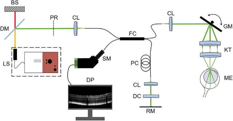
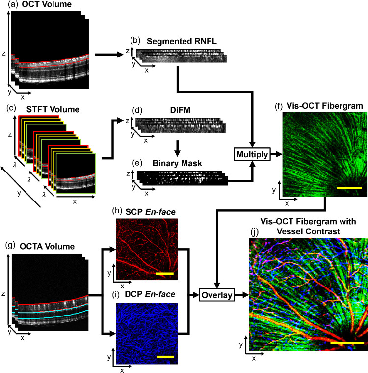
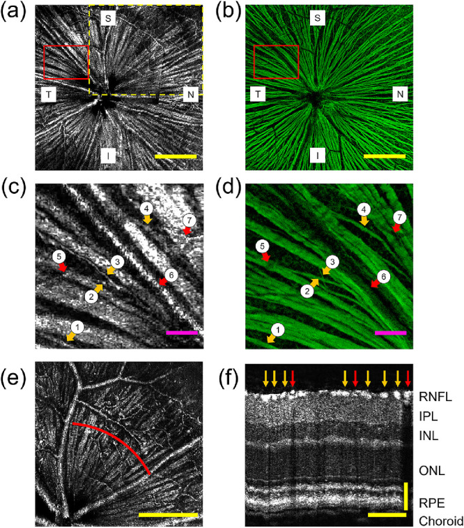
We applied visible-light optical coherence tomography (vis-OCT) to image the RGC axon bundles, referred to as vis-OCT fibergraphy, of healthy wild-type C57BL/6 mice. After vis-OCT imaging, retinas were flat-mounted, immunostained with anti-beta-III tubulin (Tuj1) antibody for RGC axons, and imaged with confocal microscopy. We quantitatively compared the RGC axon bundle networks imaged by vis-OCT and confocal microscopy using semi-log Sholl analysis.
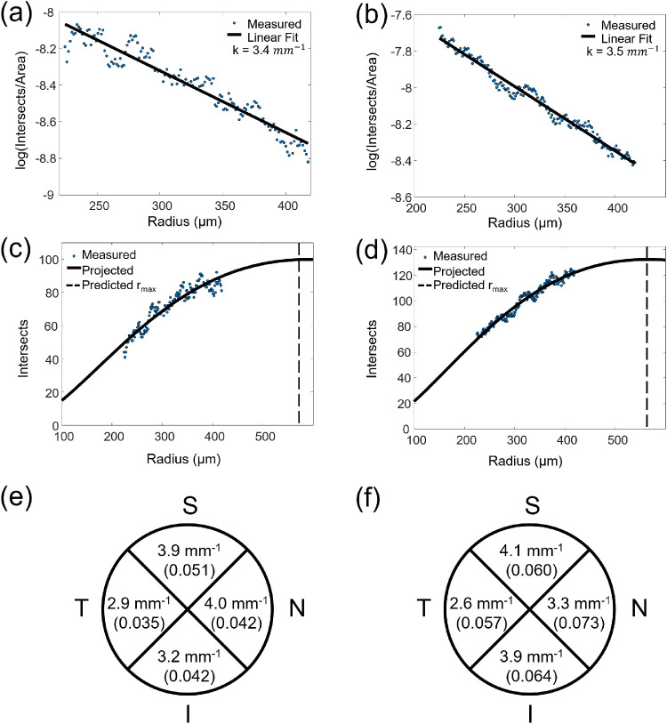
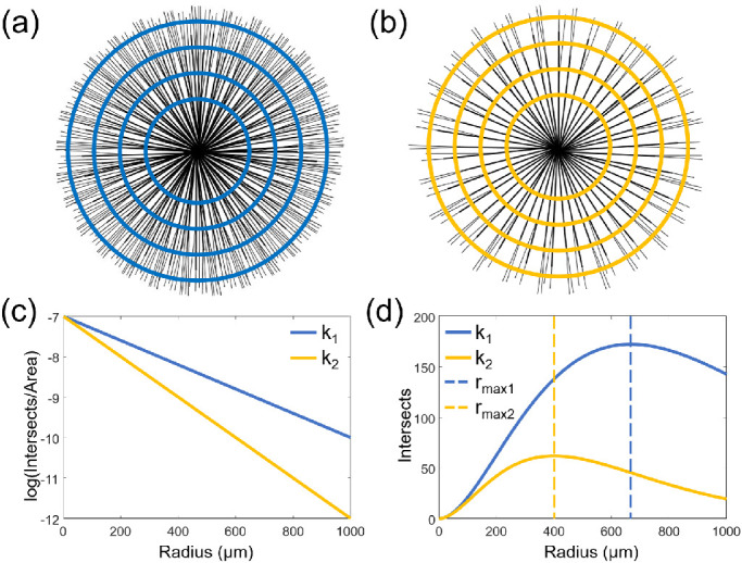
Side-by-side comparison of confocal microscopy and vis-OCT confirmed that vis-OCT fibergraphy captures true RGC axon bundle networks. The semi-log Sholl regression coefficients extracted from vis-OCT fibergrams (3.7 ± 0.8 mm) and confocal microscopy (3.6 ± 0.3 mm) images also showed good agreement with each other ( = 6).
We demonstrated the feasibility of using vis-OCT fibergraphy to visualize RGC axon bundles. Further applying Sholl analysis has the potential to identify biomarkers for non-invasively assessing RGC health.
Our novel technique for visualizing and quantifying RGC axon bundles provides a potential measurement tool for diagnosing and tracking the progression of optic neuropathies.
开发一种可视化和量化视网膜神经节细胞(RGC)轴突束的实用技术。
我们应用可见光相干断层扫描(vis-OCT)来成像健康的 C57BL/6 野生型小鼠的 RGC 轴突束,称为 vis-OCT 纤维成像。在 vis-OCT 成像后,视网膜被平展固定,用抗β-III 微管蛋白(Tuj1)抗体进行免疫染色,并用共聚焦显微镜进行成像。我们使用半对数 Sholl 分析对 vis-OCT 和共聚焦显微镜成像的 RGC 轴突束网络进行定量比较。
共聚焦显微镜和 vis-OCT 的并排比较证实,vis-OCT 纤维成像可以捕获真实的 RGC 轴突束网络。从 vis-OCT 纤维图(3.7±0.8mm)和共聚焦显微镜(3.6±0.3mm)图像中提取的半对数 Sholl 回归系数也彼此很好地吻合( = 6)。
我们证明了使用 vis-OCT 纤维成像来可视化 RGC 轴突束的可行性。进一步应用 Sholl 分析有可能识别出用于非侵入性评估 RGC 健康状况的生物标志物。
钱磊