Lamberti Giuseppe, Sisi Monia, Andrini Elisa, Palladini Arianna, Giunchi Francesca, Lollini Pier-Luigi, Ardizzoni Andrea, Gelsomino Francesco
Department of Experimental, Diagnostic and Specialty Medicine, S. Orsola-Malpighi University Hospital, Alma Mater Studiorum University of Bologna, Via Massarenti 9, 40138 Bologna, Italy.
Laboratory of Immunology and Biology of Metastasis, Department of Experimental, Diagnostic and Specialty Medicine (DIMES), University of Bologna, viale Filopanti 22, 40126 Bologna, Italy.
Cancers (Basel). 2020 Oct 26;12(11):3129. doi: 10.3390/cancers12113129.
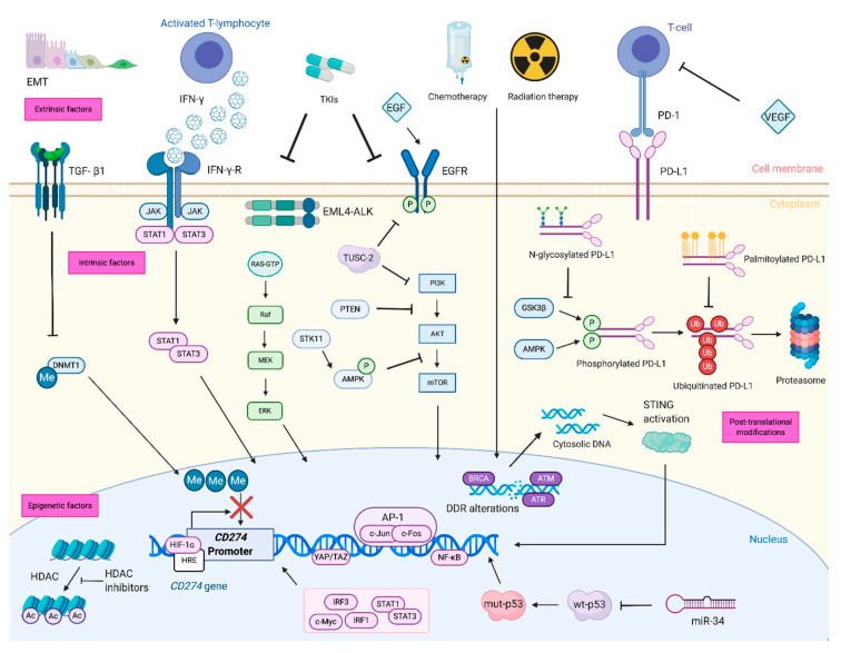
Treatment with inhibition of programmed cell death 1 (PD-1) or its ligand (PD-L1) improves survival in advanced non-small-cell lung cancer (NSCLC). Nevertheless, only a subset of patients benefit from treatment and biomarkers of response to immunotherapy are lacking. Expression of PD-L1 on tumor cells is the primary clinically-available predictive factor of response to immune checkpoint inhibitors, and its relevance in cancer immunotherapy has fostered several studies to better characterize the mechanisms that regulate PD-L1 expression. However, the factors associated with PD-L1 expression are still not well understood. Genomic alterations that activate , , and , as well as the loss of , have been associated with increased PD-L1 expression. In addition, PD-L1 expression is reported to be increased by amplification of , and decreased by deficiency. Furthermore, PD-L1 expression can be modulated by either tumor extrinsic or intrinsic factors. Among extrinsic factors, the most prominent one is interferon-γ release by immune cells, while there are several tumor intrinsic factors such as activation of the mechanistic target of rapamycin (mTOR), mitogen-activated protein kinase (MAPK) and Myc pathways that can increase PD-L1 expression. A deeper understanding of PD-L1 expression regulation is crucial for improving strategies that exploit inhibition of this immune checkpoint in the clinic, especially in NSCLC where it is central in the therapeutic algorithm. We reviewed current preclinical and clinical data about PD-L1 expression regulation in NSCLC.
抑制程序性细胞死亡1(PD-1)或其配体(PD-L1)的治疗可提高晚期非小细胞肺癌(NSCLC)患者的生存率。然而,只有一部分患者能从治疗中获益,且缺乏免疫治疗反应的生物标志物。肿瘤细胞上PD-L1的表达是免疫检查点抑制剂反应的主要临床可用预测因子,其在癌症免疫治疗中的相关性促使了多项研究,以更好地表征调节PD-L1表达的机制。然而,与PD-L1表达相关的因素仍未得到充分理解。激活、和以及缺失与PD-L1表达增加有关。此外,据报道,的扩增会增加PD-L1的表达,而的缺陷会使其降低。此外,PD-L1的表达可由肿瘤外在或内在因素调节。在外在因素中,最突出的是免疫细胞释放的干扰素-γ,而在肿瘤内在因素中,有几种因素,如雷帕霉素机制靶点(mTOR)、丝裂原活化蛋白激酶(MAPK)和Myc途径的激活,可增加PD-L1的表达。深入了解PD-L1的表达调控对于改进临床上利用这种免疫检查点抑制的策略至关重要,特别是在NSCLC中,它在治疗算法中处于核心地位。我们回顾了目前关于NSCLC中PD-L1表达调控的临床前和临床数据。