Grupo de Enfermedades Inflamatorias y Autoinmunes, Instituto de Investigación Hospital 12 de Octubre (i+12), Madrid, Spain.
Present Address: Springer Healthcare Iberica SL, Madrid, Spain.
BMC Mol Cell Biol. 2020 Oct 30;21(1):74. doi: 10.1186/s12860-020-00317-7.
The clinical efficacy of specific interleukin-6 inhibitors has confirmed the central role of IL6 in rheumatoid arthritis (RA). However the local role of IL6, in particular in synovial fibroblasts (SF) as a direct cellular target to IL6/sIL6R signal is not well characterized. The purpose of the study was to characterize the crosstalk between TNFα and IL6/sIL6R signaling to the effector pro-inflammatory response of SF.
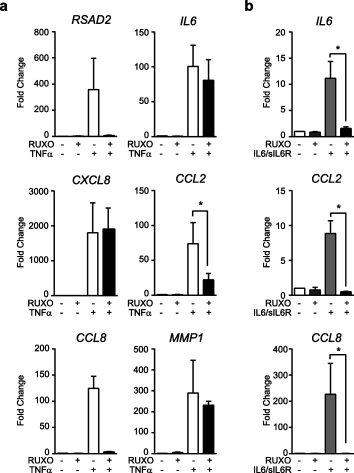
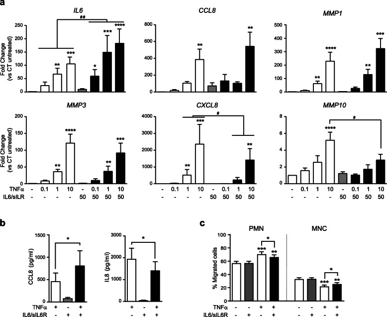
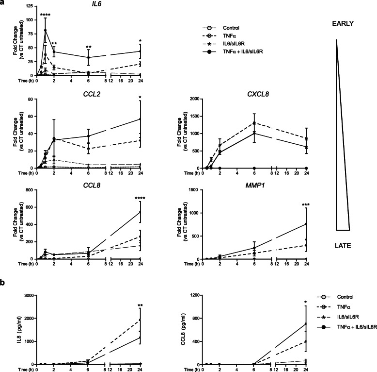
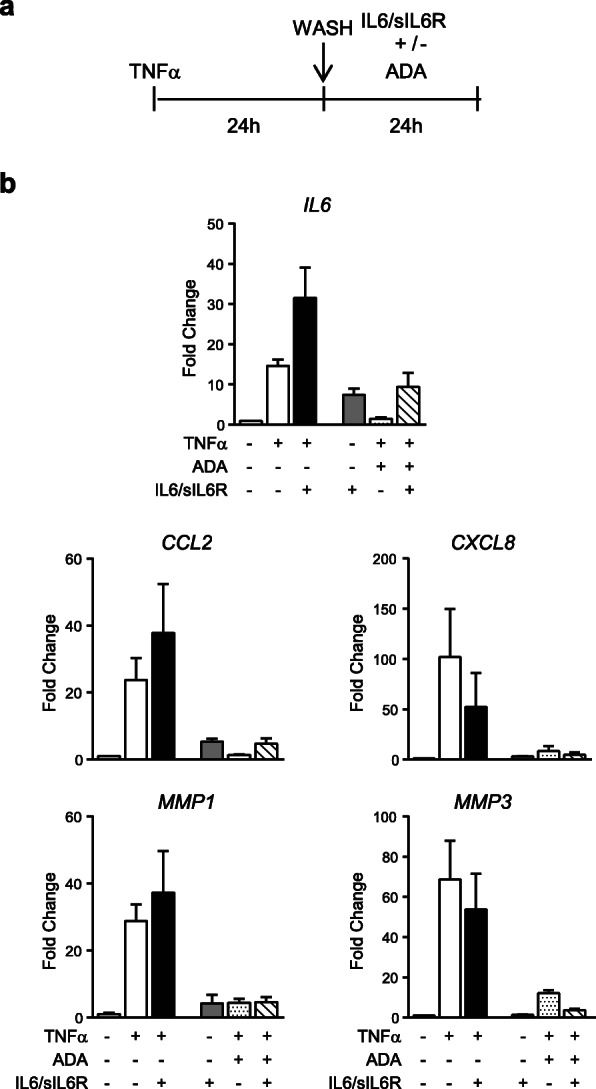
SF lines were stimulated with either TNFα, IL6/sIL6R, or both together, for the time and dose indicated for each experiment, and where indicated, cells were treated with inhibitors actinomycin D, adalimumab, ruxolitinib and cycloheximide. mRNA expression of cytokines, chemokines and matrix metalloproteases (MMPs) were analyzed by quantitative RT-PCR. Level of IL8/CXCL8 and CCL8 in culture supernatants was measured by ELISA. Mononuclear and polymorphonuclear cells migration assays were assessed by transwell using conditioned medium from SF cultures. Statistical analyses were performed as indicated in the corresponding figure legends and a p-value < 0.05 was considered statistically significant.
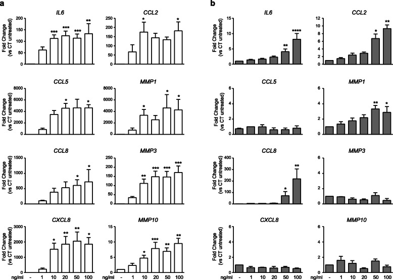
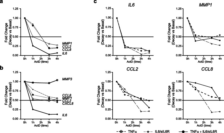
The stimulation of SF with IL6/sIL6R and TNFα, cooperatively promotes the expression of mono- and lymphocytic chemokines such as IL6, CCL8 and CCL2, as well as matrix degrading enzymes such as MMP1, while inhibiting the induction of central neutrophil chemokines such as IL8/CXCL8. These changes in the pattern of chemokines expression resulted in reduced polymorphonuclear (PMN) and increased mononuclear cells (MNC) chemoattraction by SF. Mechanistic analyses of the temporal expression of genes demonstrated that the cooperative regulation mediated by these two factors is mostly induced through de novo transcriptional mechanisms activated by IL6/sIL6R. Furthermore, we also demonstrate that TNFα and IL6/sIL6R cooperation is partially mediated by the expression of secondary factors signaling through JAK/STAT pathways.
These results point out to a highly orchestrated response to IL6 in TNFα-induced SF and provide additional insights into the role of IL6/sIL6R in the context of RA, highlighting the contribution of IL6/sIL6R to the interplay of SF with other inflammatory cells.
特定白细胞介素 6 抑制剂的临床疗效证实了白细胞介素 6(IL6)在类风湿关节炎(RA)中的核心作用。然而,IL6 在局部的作用,特别是在滑膜成纤维细胞(SF)中作为 IL6/sIL6R 信号的直接细胞靶点,尚未得到很好的描述。本研究的目的是描述 TNFα 和 IL6/sIL6R 信号之间的相互作用,以了解 SF 的效应性促炎反应。
SF 细胞系在 TNFα、IL6/sIL6R 或两者的组合下,按照每个实验规定的时间和剂量进行刺激,在有需要时,用放线菌素 D、阿达木单抗、鲁索利替尼和环已酰亚胺处理细胞。通过定量 RT-PCR 分析细胞因子、趋化因子和基质金属蛋白酶(MMPs)的 mRNA 表达。通过 ELISA 测定培养上清液中 IL8/CXCL8 和 CCL8 的水平。使用 SF 培养物的条件培养基,通过 Transwell 评估单核细胞和多形核细胞的迁移。统计分析按照相应的图例说明进行,p 值<0.05 被认为具有统计学意义。
IL6/sIL6R 和 TNFα 共同刺激 SF,协同促进单核细胞和淋巴细胞趋化因子(如 IL6、CCL8 和 CCL2)以及基质降解酶(如 MMP1)的表达,同时抑制中央中性粒细胞趋化因子(如 IL8/CXCL8)的诱导。这种趋化因子表达模式的变化导致 SF 对多形核细胞(PMN)的趋化作用降低,对单核细胞(MNC)的趋化作用增加。对基因表达的时间分析表明,这两种因素介导的协同调节主要是通过 IL6/sIL6R 激活的新的转录机制诱导的。此外,我们还证明,TNFα 和 IL6/sIL6R 的协同作用部分是通过表达通过 JAK/STAT 途径信号的二级因子来介导的。
这些结果指出了 IL6 在 TNFα 诱导的 SF 中高度协调的反应,并为 IL6/sIL6R 在 RA 背景下的作用提供了更多的见解,突出了 IL6/sIL6R 对 SF 与其他炎症细胞相互作用的贡献。