McHale Brittany, Callahan R Trey, Paras Kelsey L, Weber Martha, Kimbrell Lisa, Velázquez-Jiménez Yanet, McManamon Rita, Howerth Elizabeth W, Verocai Guilherme G
Department of Pathology, College of Veterinary Medicine, University of Georgia. 501 D.W. Brooks Drive, Athens, GA 30602, USA.
Department of Infectious Diseases, College of Veterinary Medicine, University of Georgia. 501 D.W. Brooks Drive, Athens, GA 30602, USA.
Int J Parasitol Parasites Wildl. 2020 Oct 18;13:186-190. doi: 10.1016/j.ijppaw.2020.10.005. eCollection 2020 Dec.
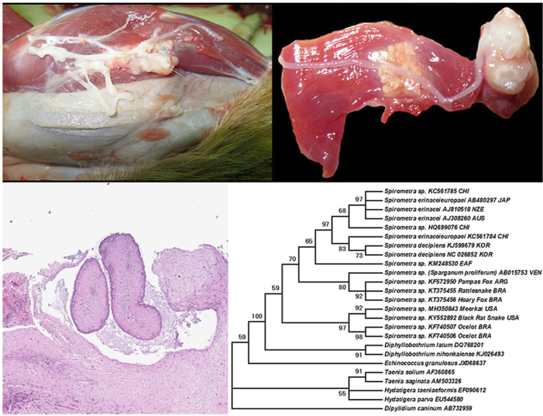
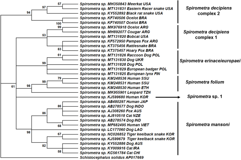
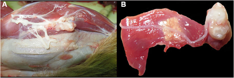
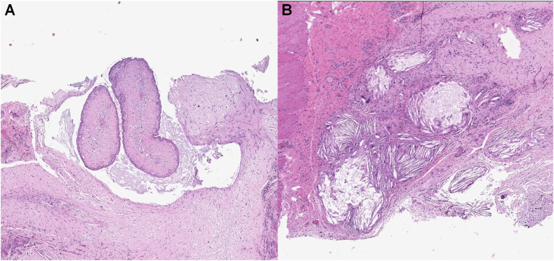
We report three cases of sparganosis due to plerocercoids of the tapeworm sp. in captive meerkats () from a zoo exhibit in the southeastern United States. Two meerkats were euthanized, one due to an uncontrollable seizure and the other due to trauma, and at necropsy cysts containing cestode larvae were observed. A third meerkat had a subcutaneous nodule surgically removed, which contained similar larvae. The third animal died years later, and had numerous cestode larvae in the pleural and peritoneal cavities. The larvae were morphologically identified as plerocercoids of diphyllobothriidean cestodes. On necropsy, multiple nodules, ranging in size from 2.5 to 3.0 cm, were observed in the subcutaneous tissue and muscles. Multifocally, separating skeletal muscle fibers were longitudinal and transversal sections of cestode larva. Histologically, parasitic cysts contained large numbers of neutrophils and macrophages, admixed with proteinaceous material. Molecular and phylogenetic analyses confirmed that specimens from one of the meerkats belonged to the genus and was closely related to plerocercoids isolated from a snake from the United States and wild felids from South America. Meerkats likely became infected by ingesting infected second intermediate hosts, such as amphibians and reptiles that may have entered the exhibit. Management practices that minimize access of meerkats and other susceptible hosts to intermediate hosts should be implemented.
我们报告了三例由裂头绦虫属裂头蚴引起的裂头蚴病病例,患病动物为美国东南部一家动物园展览中的圈养狐獴( )。两只狐獴实施了安乐死,一只因癫痫发作无法控制,另一只因外伤,尸检时观察到含有绦虫幼虫的囊肿。第三只狐獴通过手术切除了一个皮下结节,其中含有类似的幼虫。第三只动物几年后死亡,其胸腔和腹腔中有大量绦虫幼虫。这些幼虫在形态上被鉴定为双叶槽绦虫的裂头蚴。尸检时,在皮下组织和肌肉中观察到多个大小在2.5至3.0厘米之间的结节。在多处,分离的骨骼肌纤维中有绦虫幼虫的纵切面和横切面。组织学上,寄生囊肿含有大量中性粒细胞和巨噬细胞,并混有蛋白质物质。分子和系统发育分析证实,其中一只狐獴的标本属于 属,与从美国一条蛇和南美洲野生猫科动物分离出的裂头蚴密切相关。狐獴可能是通过摄食受感染的第二中间宿主而感染的,这些中间宿主可能是进入展览区域的两栖动物和爬行动物。应实施管理措施,尽量减少狐獴和其他易感宿主接触中间宿主的机会。