• 文献检索
  • 文档翻译
  • 深度研究
  • 学术资讯
  • Suppr Zotero 插件Zotero 插件
  • 邀请有礼
  • 套餐&价格
  • 历史记录
应用&插件
Suppr Zotero 插件Zotero 插件浏览器插件Mac 客户端Windows 客户端微信小程序
定价
高级版会员购买积分包购买API积分包
服务
文献检索文档翻译深度研究API 文档MCP 服务
关于我们
关于 Suppr公司介绍联系我们用户协议隐私条款
关注我们

Suppr 超能文献

核心技术专利:CN118964589B侵权必究
粤ICP备2023148730 号-1Suppr @ 2026

文献检索

告别复杂PubMed语法,用中文像聊天一样搜索,搜遍4000万医学文献。AI智能推荐,让科研检索更轻松。

立即免费搜索

文件翻译

保留排版,准确专业,支持PDF/Word/PPT等文件格式,支持 12+语言互译。

免费翻译文档

深度研究

AI帮你快速写综述,25分钟生成高质量综述,智能提取关键信息,辅助科研写作。

立即免费体验

1例前列腺癌手术及放疗后耻骨脓肿采用股直肌肌瓣治疗。

A case of pubic abscess after prostate cancer surgery and radiotherapy treated with rectus femoris muscle flap.

作者信息

Senda H, Okamoto H

机构信息

Department of Orthopaedic Surgery, Nagoya City East Medical Center, 2-23 Wakamizu 1, Chikusa, Nagoya, Aichi, 464-8547, Japan.

Department of Orthopaedic Surgery, Nagoya City University.

出版信息

JPRAS Open. 2020 Oct 6;26:54-59. doi: 10.1016/j.jpra.2020.09.006. eCollection 2020 Dec.

DOI:10.1016/j.jpra.2020.09.006
PMID:33134471
原文链接:https://pmc.ncbi.nlm.nih.gov/articles/PMC7588652/
Abstract

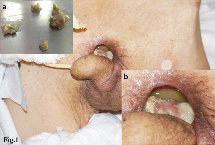
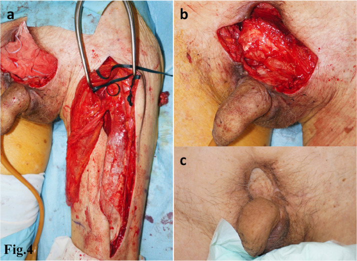
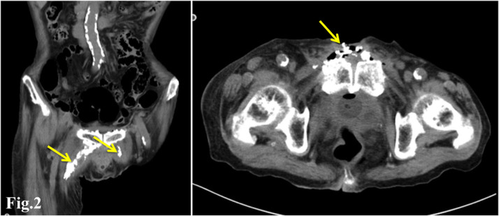
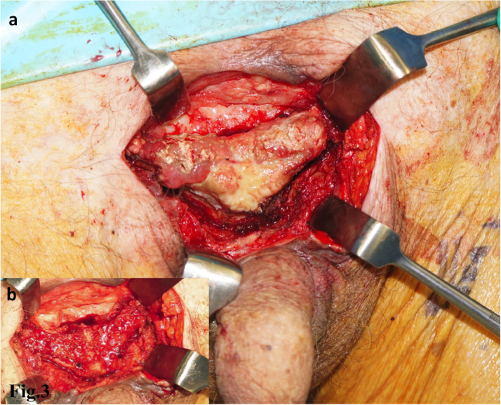
We report the case of a 95-year-old man with soft tissue deficiency associated with a pubic abscess that occurred 30 years after prostate cancer surgery and radiation therapy. A fistula with purulent discharge without any obvious cause appeared in the midline of the lower abdomen and progressed to a soft tissue defect in which several calcium phosphate stones of 5-8 mm in diameter were found. Computed tomography showed calcium deposits on the surface of the pubis and irregular zonal calcifications extending from the pubis to the medial region of both thighs. Conservative treatment did not improve the patient's condition; thus, surgical treatment was performed. The pedicled rectus femoris muscle flap was elevated from the left thigh and transferred to fill the tissue defect, then a split thickness skin graft was applied on it. The tissue defect was successfully repaired, and the patient was able to regain ambulation ability. In the present case, it was presumed that urine exudation around the bladder due to radiation cystitis was involved in the formation of ectopic calculi and subsequent infection. In reconstructing a complex defect associated with infection, using muscle flaps to fill the dead space with well vascularized tissue is considered to be appropriate. In our case, we chose a rectus femoris muscle flap, which has advantages in volume and versatility of transposition owing to long vascular pedicle and requires no microsurgical vascular anastomosis. As a result, the preoperative activity was maintained, the infection was treated, and a good course was obtained.

摘要

我们报告了一例95岁男性患者,其软组织缺损与前列腺癌手术和放射治疗30年后发生的耻骨脓肿有关。下腹中线出现了一个无明显原因的脓性分泌物瘘管,并发展为软组织缺损,在其中发现了数颗直径为5-8毫米的磷酸钙结石。计算机断层扫描显示耻骨表面有钙沉积,以及从耻骨延伸至双侧大腿内侧区域的不规则带状钙化。保守治疗未能改善患者病情,因此进行了手术治疗。从左大腿掀起带蒂股直肌肌瓣并转移以填充组织缺损,然后在其上应用中厚皮片移植。组织缺损成功修复,患者能够恢复行走能力。在本病例中,推测放射性膀胱炎导致膀胱周围尿液渗出与异位结石的形成及随后的感染有关。在重建与感染相关的复杂缺损时,使用肌瓣用血管丰富的组织填充死腔被认为是合适的。在我们的病例中,我们选择了股直肌肌瓣,由于其血管蒂长,在体积和转位的通用性方面具有优势,且无需显微外科血管吻合。结果,患者术前的活动能力得以维持,感染得到治疗,病情进展良好。

https://cdn.ncbi.nlm.nih.gov/pmc/blobs/5288/7588652/77d306d6e4bd/gr4.jpg
https://cdn.ncbi.nlm.nih.gov/pmc/blobs/5288/7588652/6ba064454597/gr1.jpg
https://cdn.ncbi.nlm.nih.gov/pmc/blobs/5288/7588652/8edbe57d6cfa/gr2.jpg
https://cdn.ncbi.nlm.nih.gov/pmc/blobs/5288/7588652/c348f895f467/gr3.jpg
https://cdn.ncbi.nlm.nih.gov/pmc/blobs/5288/7588652/77d306d6e4bd/gr4.jpg
https://cdn.ncbi.nlm.nih.gov/pmc/blobs/5288/7588652/6ba064454597/gr1.jpg
https://cdn.ncbi.nlm.nih.gov/pmc/blobs/5288/7588652/8edbe57d6cfa/gr2.jpg
https://cdn.ncbi.nlm.nih.gov/pmc/blobs/5288/7588652/c348f895f467/gr3.jpg
https://cdn.ncbi.nlm.nih.gov/pmc/blobs/5288/7588652/77d306d6e4bd/gr4.jpg

相似文献

1
A case of pubic abscess after prostate cancer surgery and radiotherapy treated with rectus femoris muscle flap.1例前列腺癌手术及放疗后耻骨脓肿采用股直肌肌瓣治疗。
JPRAS Open. 2020 Oct 6;26:54-59. doi: 10.1016/j.jpra.2020.09.006. eCollection 2020 Dec.
2
Use of Rectus Femoris Muscle Flap in Patients With Absent Profunda Femoris Vascular Flow.股深血管血流缺失患者中股直肌肌瓣的应用
Eplasty. 2022 Sep 15;22:e42. eCollection 2022.
3
Distally based anteromedial thigh flaps pedicled on the rectus femoris branch of the lateral circumflex femoral artery for reconstruction of soft-tissue defect of the knee.以旋股外侧动脉降支为蒂的股前内收肌肌皮瓣游离移植修复膝关节软组织缺损
J Plast Reconstr Aesthet Surg. 2018 May;71(5):743-749. doi: 10.1016/j.bjps.2018.01.007. Epub 2018 Feb 7.
4
[DESIGN AND CLINICAL APPLICATIONS OF DISTALLY BASED ANTEROMEDIAL THIGH FLAPS PEDICLED WITH PERFORATING VESSELS FROM RECTUS FEMORIS BRANCH].[以股直肌分支穿支血管为蒂的远端蒂股前内侧皮瓣的设计与临床应用]
Zhongguo Xiu Fu Chong Jian Wai Ke Za Zhi. 2016 Dec 8;30(12):1502-1506. doi: 10.7507/1002-1892.20160311.
5
Management of complex groin wounds: preferred use of the rectus femoris muscle flap.复杂腹股沟伤口的处理:股直肌肌瓣的优先应用
Plast Reconstr Surg. 2005 Mar;115(3):776-83; discussion 784-5. doi: 10.1097/01.prs.0000152436.50604.04.
6
[Repairing postoperative soft tissue defects of tibia and ankle open fractures with muscle flap pedicled with medial half of soleus].[比目鱼肌内侧半肌瓣修复胫腓骨及踝关节开放性骨折术后软组织缺损]
Zhongguo Xiu Fu Chong Jian Wai Ke Za Zhi. 2009 Dec;23(12):1440-2.
7
Revisiting vascular supply of the rectus femoris and its relevance in the harvest of the anterolateral thigh flap.重新审视股直肌的血管供应及其在股前外侧皮瓣切取中的相关性。
Ann Plast Surg. 2013 Nov;71(5):586-90. doi: 10.1097/SAP.0b013e318255a2ac.
8
[Pedicled anterolateral thigh flap reconstruction in the region of the lower abdomen, groin, perineum, and hip].带蒂股前外侧皮瓣在下腹部、腹股沟、会阴及臀部区域的重建
Oper Orthop Traumatol. 2022 Oct;34(5):372-378. doi: 10.1007/s00064-022-00772-9. Epub 2022 May 29.
9
[Combination mode and optimization strategy of harvest procedure of anterolateral thigh chimeric perforator myocutaneous flap].股前外侧嵌合穿支肌皮瓣切取程序的组合模式与优化策略
Zhongguo Xiu Fu Chong Jian Wai Ke Za Zhi. 2023 Feb 15;37(2):180-184. doi: 10.7507/1002-1892.202209059.
10
[Clinical effects of retrograde anterolateral thigh perforator flaps assisted with computed tomography angiography in repairing skin and soft tissue defects around the knee or in proximal lower leg].计算机断层血管造影辅助逆行股前外侧穿支皮瓣修复膝关节周围及小腿近端皮肤软组织缺损的临床效果
Zhonghua Shao Shang Za Zhi. 2021 Apr 20;37(4):356-362. doi: 10.3760/cma.j.cn501120-20200905-00401.

本文引用的文献

1
[Collection in the thigh revealing bladder fistula after radiotherapy for prostate cancer].[前列腺癌放疗后大腿部积液显示膀胱瘘]
Pan Afr Med J. 2018 Jul 5;30:197. doi: 10.11604/pamj.2018.30.197.16037. eCollection 2018.
2
Radiotherapy-related complications presenting to a urology department: a more common problem than previously thought?泌尿科就诊的放射治疗相关并发症:比以往想象的更常见的问题?
BJU Int. 2018 May;121 Suppl 3:28-32. doi: 10.1111/bju.14145. Epub 2018 Feb 27.
3
Pubic Bone Osteomyelitis and Pubosymphyseal Urinary Fistula: A Poorly Recognized Complication in Prostate Cancer Survivors.
耻骨骨髓炎和耻骨联合尿道瘘:前列腺癌幸存者中一种未被充分认识的并发症。
Oncology (Williston Park). 2017 Mar 15;31(3):169-73.
4
Management of Radiation Anterior Prostato-symphyseal Fistulas With Interposition Rectus Abdominis Muscle Flap.应用腹直肌肌瓣修复放射性前列腺-耻骨联合前瘘的治疗
Urology. 2016 Jun;92:122-6. doi: 10.1016/j.urology.2016.01.029. Epub 2016 Feb 12.
5
Radiation Induced Cystitis and Proctitis - Prediction, Assessment and Management.放射性膀胱炎和直肠炎——预测、评估与管理
Asian Pac J Cancer Prev. 2015;16(14):5589-94. doi: 10.7314/apjcp.2015.16.14.5589.
6
The treatment of chronic osteomyelitis: a 10 year audit.慢性骨髓炎的治疗:一项为期10年的审计。
J Plast Reconstr Aesthet Surg. 2006;59(1):11-5. doi: 10.1016/j.bjps.2005.07.002.
7
Abscess formation in the prevesical space and bilateral thigh muscles secondary to osteomyelitis of the pubis--basis of the anatomy between the prevesical space and femoral sheath.耻骨骨髓炎继发膀胱前间隙及双侧大腿肌肉脓肿形成——膀胱前间隙与股鞘之间的解剖学基础
Scand J Urol Nephrol. 2004;38(5):440-1. doi: 10.1080/00365590410031652.
8
An algorithm for abdominal wall reconstruction.一种腹壁重建算法。
Plast Reconstr Surg. 2000 Jan;105(1):202-16; quiz 217. doi: 10.1097/00006534-200001000-00036.
9
Spontaneous rupture of the urinary bladder is not a rare complication of radiotherapy for cervical cancer: report of six cases.膀胱自发性破裂并非宫颈癌放疗的罕见并发症:6例报告
Gynecol Oncol. 1999 Jun;73(3):439-42. doi: 10.1006/gyno.1999.5347.
10
Experimental evaluation of the antibiotic carrying capacity of a muscle flap into a fibrotic cavity.
Plast Reconstr Surg. 1988 Feb;81(2):162-70. doi: 10.1097/00006534-198802000-00003.