The George & Anne Ryan Institute for Neuroscience, University of Rhode Island, 130 Flagg Road, Kingston, RI, 02881, United States.
Department of Biomedical and Pharmaceutical Sciences, College of Pharmacy, University of Rhode Island, Kingston, RI, 02881, USA.
Cell Mol Neurobiol. 2022 May;42(4):985-996. doi: 10.1007/s10571-020-00987-z. Epub 2020 Nov 2.
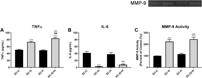
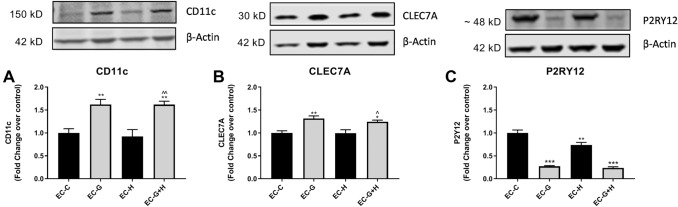
Diabetes is strongly linked to the development of Alzheimer's disease (AD), though the mechanisms for this enhanced risk are unclear. Because vascular inflammation is a consistent feature of both diabetes and AD, the cerebral microcirculation could be a key target for the effects of diabetes in the brain. The goal of this study is to explore whether brain endothelial cells, injured by diabetes-related insults, glucose and hypoxia, can affect inflammatory and activation processes in microglia in vitro. Human brain microvascular endothelial cells (HBMVECs) were either treated with 5 mM glucose (control), 30 mM glucose (high glucose), exposed to hypoxia, or exposed to hypoxia plus high glucose. HBMVEC-conditioned medium was then used to treat BV-2 microglia. Alterations in microglia phenotype were assessed through measurement of nitric oxide (NO), cytokine production, microglial activation state markers, and microglial phagocytosis. HBMVECs were injured by exposure to glucose and/or hypoxia, as assessed by release of LDH, interleukin (IL)-1β, and reactive oxygen species (ROS). HBMVECs injured by glucose and hypoxia induced increases in microglial production of NO, tumor necrosis factor-α (TNFα) and matrix metalloproteinase (MMP)-9. Injured HBMVECs significantly increased microglial expression of CD11c and CLEC7A, and decreased expression of the homeostatic marker P2RY12. Finally, bead uptake by BV-2 cells, an index of phagocytic ability, was elevated by conditioned media from injured HBMVECs. The demonstration that injury to brain endothelial cells by diabetic-associated insults, glucose and hypoxia, promotes microglial inflammation supports the idea that the cerebral microcirculation is a critical locus for the deleterious effects of diabetes in the AD brain.
糖尿病与阿尔茨海默病(AD)的发展密切相关,但这种风险增加的机制尚不清楚。由于血管炎症是糖尿病和 AD 的共同特征,因此大脑微循环可能是糖尿病对大脑影响的关键靶点。本研究旨在探讨糖尿病相关损伤、葡萄糖和缺氧是否会影响体外脑内皮细胞中炎症和激活过程。将人脑微血管内皮细胞(HBMVEC)分别用 5 mM 葡萄糖(对照)、30 mM 葡萄糖(高糖)、缺氧或缺氧加高糖处理。然后用 HBMVEC 条件培养基处理 BV-2 小胶质细胞。通过测量一氧化氮(NO)、细胞因子产生、小胶质细胞激活状态标志物和小胶质细胞吞噬作用来评估小胶质细胞表型的变化。通过释放乳酸脱氢酶(LDH)、白细胞介素(IL)-1β 和活性氧(ROS)来评估 HBMVEC 暴露于葡萄糖和/或缺氧后的损伤情况。葡萄糖和缺氧损伤的 HBMVEC 诱导小胶质细胞产生的一氧化氮(NO)、肿瘤坏死因子-α(TNFα)和基质金属蛋白酶(MMP)-9 增加。受损的 HBMVEC 显著增加了小胶质细胞中 CD11c 和 CLEC7A 的表达,并降低了稳态标志物 P2RY12 的表达。最后,由受损 HBMVEC 的条件培养基引起的 BV-2 细胞珠摄取增加,这是吞噬能力的一个指标。糖尿病相关损伤、葡萄糖和缺氧对脑内皮细胞的损伤促进小胶质细胞炎症的证据支持这样一种观点,即大脑微循环是糖尿病对 AD 大脑有害影响的关键部位。