• 文献检索
  • 文档翻译
  • 深度研究
  • 学术资讯
  • Suppr Zotero 插件Zotero 插件
  • 邀请有礼
  • 套餐&价格
  • 历史记录
应用&插件
Suppr Zotero 插件Zotero 插件浏览器插件Mac 客户端Windows 客户端微信小程序
定价
高级版会员购买积分包购买API积分包
服务
文献检索文档翻译深度研究API 文档MCP 服务
关于我们
关于 Suppr公司介绍联系我们用户协议隐私条款
关注我们

Suppr 超能文献

核心技术专利:CN118964589B侵权必究
粤ICP备2023148730 号-1Suppr @ 2026

文献检索

告别复杂PubMed语法,用中文像聊天一样搜索,搜遍4000万医学文献。AI智能推荐,让科研检索更轻松。

立即免费搜索

文件翻译

保留排版,准确专业,支持PDF/Word/PPT等文件格式,支持 12+语言互译。

免费翻译文档

深度研究

AI帮你快速写综述,25分钟生成高质量综述,智能提取关键信息,辅助科研写作。

立即免费体验

同时敲除 L 型 Ca1.3(α)和 T 型 Ca3.1(α)钙通道基因会破坏心脏自律性。

Concomitant genetic ablation of L-type Ca1.3 (α) and T-type Ca3.1 (α) Ca channels disrupts heart automaticity.

机构信息

Institut de Génomique Fonctionnelle, Université de Montpellier, CNRS, Inserm, 141, rue de la cardonille, 34094, Montpellier, France.

LabEx ICST, Montpellier, France.

出版信息

Sci Rep. 2020 Nov 3;10(1):18906. doi: 10.1038/s41598-020-76049-7.

DOI:10.1038/s41598-020-76049-7
PMID:33144668
原文链接:https://pmc.ncbi.nlm.nih.gov/articles/PMC7642305/
Abstract

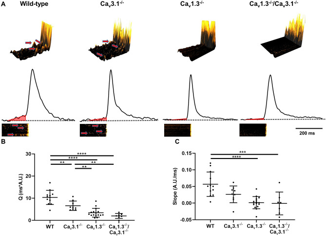
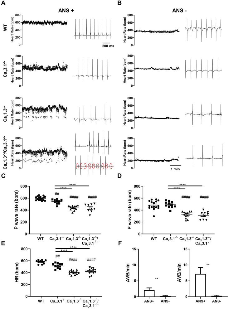
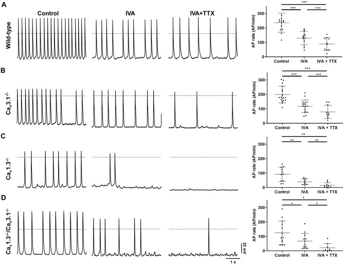
Cardiac automaticity is set by pacemaker activity of the sinus node (SAN). In addition to the ubiquitously expressed cardiac voltage-gated L-type Ca1.2 Ca channel isoform, pacemaker cells within the SAN and the atrioventricular node co-express voltage-gated L-type Ca1.3 and T-type Ca3.1 Ca channels (SAN-VGCCs). The role of SAN-VGCCs in automaticity is incompletely understood. We used knockout mice carrying individual genetic ablation of Ca1.3 (Ca1.3) or Ca3.1 (Ca3.1) channels and double mutant Ca1.3/Ca3.1 mice expressing only Ca1.2 channels. We show that concomitant loss of SAN-VGCCs prevents physiological SAN automaticity, blocks impulse conduction and compromises ventricular rhythmicity. Coexpression of SAN-VGCCs is necessary for impulse formation in the central SAN. In mice lacking SAN-VGCCs, residual pacemaker activity is predominantly generated in peripheral nodal and extranodal sites by f-channels and TTX-sensitive Na channels. In beating SAN cells, ablation of SAN-VGCCs disrupted late diastolic local intracellular Ca release, which demonstrates an important role for these channels in supporting the sarcoplasmic reticulum based "Ca clock" mechanism during normal pacemaking. These data implicate an underappreciated role for co-expression of SAN-VGCCs in heart automaticity and define an integral role for these channels in mechanisms that control the heartbeat.

摘要

心脏自律性由窦房结(SAN)的起搏器活动设定。除了广泛表达的心脏电压门控 L 型 Ca1.2 Ca 通道同工型外,SAN 和房室结内的起搏细胞还共同表达电压门控 L 型 Ca1.3 和 T 型 Ca3.1 Ca 通道(SAN-VGCCs)。SAN-VGCCs 在自律性中的作用尚未完全了解。我们使用携带单个 Ca1.3(Ca1.3)或 Ca3.1(Ca3.1)通道基因缺失的敲除小鼠和仅表达 Ca1.2 通道的 Ca1.3/Ca3.1 双突变小鼠。我们表明,SAN-VGCCs 的同时缺失可防止生理 SAN 自律性,阻断冲动传导并损害心室节律性。SAN-VGCCs 的共表达对于 SAN 中央冲动形成是必需的。在缺乏 SAN-VGCCs 的小鼠中,残余的起搏活性主要由 f 通道和 TTX 敏感的 Na 通道在周围节段和节外部位产生。在跳动的 SAN 细胞中,SAN-VGCCs 的消融破坏了舒张晚期局部细胞内 Ca 释放,这表明这些通道在支持正常起搏时基于肌浆网的“Ca 钟”机制中发挥重要作用。这些数据表明 SAN-VGCCs 的共表达在心脏自律性中具有被低估的作用,并定义了这些通道在控制心跳的机制中的不可或缺的作用。

https://cdn.ncbi.nlm.nih.gov/pmc/blobs/7fde/7642305/c5046f2f76c6/41598_2020_76049_Fig9_HTML.jpg
https://cdn.ncbi.nlm.nih.gov/pmc/blobs/7fde/7642305/b1944eebbd32/41598_2020_76049_Fig1_HTML.jpg
https://cdn.ncbi.nlm.nih.gov/pmc/blobs/7fde/7642305/1b5b3b2a237c/41598_2020_76049_Fig2_HTML.jpg
https://cdn.ncbi.nlm.nih.gov/pmc/blobs/7fde/7642305/ab933c18b099/41598_2020_76049_Fig3_HTML.jpg
https://cdn.ncbi.nlm.nih.gov/pmc/blobs/7fde/7642305/219ea8643441/41598_2020_76049_Fig4_HTML.jpg
https://cdn.ncbi.nlm.nih.gov/pmc/blobs/7fde/7642305/284dc79ce145/41598_2020_76049_Fig5_HTML.jpg
https://cdn.ncbi.nlm.nih.gov/pmc/blobs/7fde/7642305/25b9163338d4/41598_2020_76049_Fig6_HTML.jpg
https://cdn.ncbi.nlm.nih.gov/pmc/blobs/7fde/7642305/46a0552d95c9/41598_2020_76049_Fig7_HTML.jpg
https://cdn.ncbi.nlm.nih.gov/pmc/blobs/7fde/7642305/a9c8986099bb/41598_2020_76049_Fig8_HTML.jpg
https://cdn.ncbi.nlm.nih.gov/pmc/blobs/7fde/7642305/c5046f2f76c6/41598_2020_76049_Fig9_HTML.jpg

相似文献

1
Concomitant genetic ablation of L-type Ca1.3 (α) and T-type Ca3.1 (α) Ca channels disrupts heart automaticity.同时敲除 L 型 Ca1.3(α)和 T 型 Ca3.1(α)钙通道基因会破坏心脏自律性。
Sci Rep. 2020 Nov 3;10(1):18906. doi: 10.1038/s41598-020-76049-7.
2
Channelopathies of voltage-gated L-type Cav1.3/α and T-type Cav3.1/α Ca channels in dysfunction of heart automaticity.电压门控 L 型 Cav1.3/α 和 T 型 Cav3.1/α Ca 通道通道病在心自动节律功能障碍中的作用。
Pflugers Arch. 2020 Jul;472(7):817-830. doi: 10.1007/s00424-020-02421-1. Epub 2020 Jun 29.
3
Functional role of voltage gated Ca(2+) channels in heart automaticity.电压门控Ca(2+)通道在心脏自律性中的功能作用。
Front Physiol. 2015 Feb 2;6:19. doi: 10.3389/fphys.2015.00019. eCollection 2015.
4
L-type Cav1.3 channels regulate ryanodine receptor-dependent Ca2+ release during sino-atrial node pacemaker activity.L型Cav1.3通道在窦房结起搏活动期间调节兰尼碱受体依赖性Ca2+释放。
Cardiovasc Res. 2016 Mar 1;109(3):451-61. doi: 10.1093/cvr/cvw006. Epub 2016 Jan 19.
5
Bradycardia and slowing of the atrioventricular conduction in mice lacking CaV3.1/alpha1G T-type calcium channels.缺乏CaV3.1/α1G T型钙通道的小鼠出现心动过缓和房室传导减慢。
Circ Res. 2006 Jun 9;98(11):1422-30. doi: 10.1161/01.RES.0000225862.14314.49. Epub 2006 May 11.
6
T-type calcium current contributes to escape automaticity and governs the occurrence of lethal arrhythmias after atrioventricular block in mice.T 型钙电流有助于逃避自动性,并控制房室传导阻滞后小鼠致命性心律失常的发生。
Circ Arrhythm Electrophysiol. 2013 Aug;6(4):799-808. doi: 10.1161/CIRCEP.113.000407. Epub 2013 Jul 24.
7
T-type channels in the sino-atrial and atrioventricular pacemaker mechanism.窦房结和房室结起搏机制中的T型通道。
Pflugers Arch. 2014 Apr;466(4):791-9. doi: 10.1007/s00424-014-1482-6. Epub 2014 Feb 27.
8
Inhibition of G protein-gated K channels by tertiapin-Q rescues sinus node dysfunction and atrioventricular conduction in mouse models of primary bradycardia.三磷酸哌喹抑制 G 蛋白门控钾通道可挽救原发性心动过缓小鼠模型中的窦房结功能障碍和房室传导阻滞。
Sci Rep. 2020 Jun 17;10(1):9835. doi: 10.1038/s41598-020-66673-8.
9
Functional roles of Ca(v)1.3, Ca(v)3.1 and HCN channels in automaticity of mouse atrioventricular cells: insights into the atrioventricular pacemaker mechanism.钙通道(Ca(v)1.3、Ca(v)3.1)和 HCN 通道在小鼠房室细胞自律性中的功能作用:房室起搏机制的研究进展。
Channels (Austin). 2011 May-Jun;5(3):251-61. doi: 10.4161/chan.5.3.15266. Epub 2011 May 1.
10
Ca1.3 L-type Ca channel contributes to the heartbeat by generating a dihydropyridine-sensitive persistent Na current.钙通道 1.3 L 型钙通道通过产生二氢吡啶敏感的持续钠电流来促进心跳。
Sci Rep. 2017 Aug 11;7(1):7869. doi: 10.1038/s41598-017-08191-8.

引用本文的文献

1
Electrophysiological Mechanisms and Therapeutic Potential of Calcium Channels in Atrial Fibrillation.心房颤动中钙通道的电生理机制及治疗潜力
Rev Cardiovasc Med. 2025 Jun 25;26(6):33507. doi: 10.31083/RCM33507. eCollection 2025 Jun.
2
Role of Cav1.3 Channels in Brain-Heart Interactions: An Unexpected Journey.Cav1.3通道在脑-心相互作用中的作用:一段意想不到的历程。
Biomedicines. 2025 Jun 4;13(6):1376. doi: 10.3390/biomedicines13061376.
3
Aging reduces the number and function of L-type calcium channels in the membrane of cardiac pacemaker cells.

本文引用的文献

1
Synchronized Cardiac Impulses Emerge From Heterogeneous Local Calcium Signals Within and Among Cells of Pacemaker Tissue.起搏组织细胞内和细胞间的异质局部钙信号中出现同步心脏冲动。
JACC Clin Electrophysiol. 2020 Aug;6(8):907-931. doi: 10.1016/j.jacep.2020.06.022.
2
Impaired neuronal sodium channels cause intranodal conduction failure and reentrant arrhythmias in human sinoatrial node.神经元钠通道功能障碍导致人心房结内传导阻滞和折返性心律失常。
Nat Commun. 2020 Jan 24;11(1):512. doi: 10.1038/s41467-019-14039-8.
3
Identification of CACNA1D variants associated with sinoatrial node dysfunction and deafness in additional Pakistani families reveals a clinical significance.
衰老会减少心脏起搏器细胞膜上L型钙通道的数量和功能。
bioRxiv. 2025 May 27:2022.06.22.497267. doi: 10.1101/2022.06.22.497267.
4
The Neglected Internodal Tract-A Cardiac Conduction System Structure Homologous to the Development and Regulation of the Sinoatrial Node.被忽视的结间束——一种与窦房结发育和调节同源的心脏传导系统结构
Rev Cardiovasc Med. 2025 Apr 22;26(4):27882. doi: 10.31083/RCM27882. eCollection 2025 Apr.
5
CACNA1G, A Heterotaxy Candidate Gene, Plays a Role in Ciliogenesis and Left-Right Patterning in Xenopus tropicalis.CACNA1G,一个心脏异位候选基因,在热带爪蟾的纤毛发生和左右模式形成中发挥作用。
Genesis. 2025 Feb;63(1):e70009. doi: 10.1002/dvg.70009.
6
The electrophysiological effects of Tongyang Huoxue granules on the ignition phase during hypoxia/reoxygenation injury in sinoatrial node cells.通阳活血颗粒对窦房结细胞缺氧/复氧损伤时触发期的电生理效应。
Front Physiol. 2024 Jun 7;15:1402478. doi: 10.3389/fphys.2024.1402478. eCollection 2024.
7
Cardiac L-type calcium channel regulation by Leucine-Rich Repeat-Containing Protein 10.富含亮氨酸重复蛋白 10 对心脏 L 型钙通道的调节作用。
Channels (Austin). 2024 Dec;18(1):2355121. doi: 10.1080/19336950.2024.2355121. Epub 2024 May 19.
8
Pacemaker Channels and the Chronotropic Response in Health and Disease.起搏器通道与健康和疾病中的变时性反应。
Circ Res. 2024 May 10;134(10):1348-1378. doi: 10.1161/CIRCRESAHA.123.323250. Epub 2024 May 9.
9
Selective blockade of Ca1.2 (α1C) versus Ca1.3 (α1D) L-type calcium channels by the black mamba toxin calciseptine.黑曼巴毒素 calciseptine 对 Ca1.2(α1C)与 Ca1.3(α1D)L 型钙通道的选择性阻断。
Nat Commun. 2024 Jan 2;15(1):54. doi: 10.1038/s41467-023-43502-w.
10
Biophysical properties of Na1.5 channels from atrial-like and ventricular-like cardiomyocytes derived from human induced pluripotent stem cells.人诱导多能干细胞来源的心房样和心室样心肌细胞中的 Na1.5 通道的生物物理特性。
Sci Rep. 2023 Nov 24;13(1):20685. doi: 10.1038/s41598-023-47310-6.
在其他巴基斯坦家庭中鉴定与窦房结功能障碍和耳聋相关的 CACNA1D 变体,揭示了其临床意义。
J Hum Genet. 2019 Feb;64(2):153-160. doi: 10.1038/s10038-018-0542-8. Epub 2018 Nov 30.
4
Positive Feedback Mechanisms among Local Ca Releases, NCX, and I Ignite Pacemaker Action Potentials.局部钙释放、钠钙交换体(NCX)和I之间的正反馈机制引发起搏器动作电位。
Biophys J. 2018 Apr 24;114(8):2024. doi: 10.1016/j.bpj.2018.03.024.
5
Increasing T-type calcium channel activity by β-adrenergic stimulation contributes to β-adrenergic regulation of heart rates.β-肾上腺素能刺激增加 T 型钙通道活性有助于β-肾上腺素能调节心率。
J Physiol. 2018 Apr 1;596(7):1137-1151. doi: 10.1113/JP274756. Epub 2018 Jan 24.
6
Ca1.3 L-type Ca channel contributes to the heartbeat by generating a dihydropyridine-sensitive persistent Na current.钙通道 1.3 L 型钙通道通过产生二氢吡啶敏感的持续钠电流来促进心跳。
Sci Rep. 2017 Aug 11;7(1):7869. doi: 10.1038/s41598-017-08191-8.
7
Autoimmune channelopathies as a novel mechanism in cardiac arrhythmias.自身免疫性通道病作为心律失常的一种新机制。
Nat Rev Cardiol. 2017 Sep;14(9):521-535. doi: 10.1038/nrcardio.2017.61. Epub 2017 May 4.
8
G protein-gated IKACh channels as therapeutic targets for treatment of sick sinus syndrome and heart block.G蛋白门控内向整流钾通道作为治疗病态窦房结综合征和心脏传导阻滞的治疗靶点。
Proc Natl Acad Sci U S A. 2016 Feb 16;113(7):E932-41. doi: 10.1073/pnas.1517181113. Epub 2016 Feb 1.
9
L-type Cav1.3 channels regulate ryanodine receptor-dependent Ca2+ release during sino-atrial node pacemaker activity.L型Cav1.3通道在窦房结起搏活动期间调节兰尼碱受体依赖性Ca2+释放。
Cardiovasc Res. 2016 Mar 1;109(3):451-61. doi: 10.1093/cvr/cvw006. Epub 2016 Jan 19.
10
Burst pacemaker activity of the sinoatrial node in sodium-calcium exchanger knockout mice.钠钙交换体基因敲除小鼠窦房结的爆发式起搏器活动
Proc Natl Acad Sci U S A. 2015 Aug 4;112(31):9769-74. doi: 10.1073/pnas.1505670112. Epub 2015 Jul 20.