• 文献检索
  • 文档翻译
  • 深度研究
  • 学术资讯
  • Suppr Zotero 插件Zotero 插件
  • 邀请有礼
  • 套餐&价格
  • 历史记录
应用&插件
Suppr Zotero 插件Zotero 插件浏览器插件Mac 客户端Windows 客户端微信小程序
定价
高级版会员购买积分包购买API积分包
服务
文献检索文档翻译深度研究API 文档MCP 服务
关于我们
关于 Suppr公司介绍联系我们用户协议隐私条款
关注我们

Suppr 超能文献

核心技术专利:CN118964589B侵权必究
粤ICP备2023148730 号-1Suppr @ 2026

文献检索

告别复杂PubMed语法,用中文像聊天一样搜索,搜遍4000万医学文献。AI智能推荐,让科研检索更轻松。

立即免费搜索

文件翻译

保留排版,准确专业,支持PDF/Word/PPT等文件格式,支持 12+语言互译。

免费翻译文档

深度研究

AI帮你快速写综述,25分钟生成高质量综述,智能提取关键信息,辅助科研写作。

立即免费体验

利用脉络膜上腔开口最小 rim 宽度和 RNFL 对早期正常眼压性青光眼和青光眼疑似患者进行深度学习分类。

Deep learning classification of early normal-tension glaucoma and glaucoma suspects using Bruch's membrane opening-minimum rim width and RNFL.

机构信息

Department of Mathematics Education, School of Education, Kyungnam University, Changwon, Republic of Korea.

Department of Ophthalmology, Gyeongsang National University Changwon Hospital, Gyeongsang National University, School of Medicine, 11 Samjeongja-ro, Seongsan-gu, Changwon, Gyeongsangnam-do, 51472, Republic of Korea.

出版信息

Sci Rep. 2020 Nov 4;10(1):19042. doi: 10.1038/s41598-020-76154-7.

DOI:10.1038/s41598-020-76154-7
PMID:33149191
原文链接:https://pmc.ncbi.nlm.nih.gov/articles/PMC7643070/
Abstract

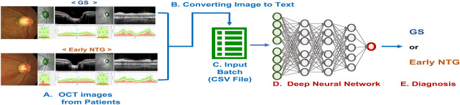
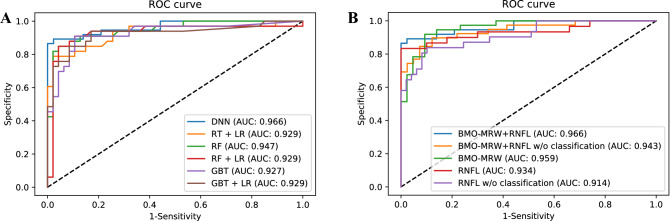
We aimed to classify early normal-tension glaucoma (NTG) and glaucoma suspect (GS) using Bruch's membrane opening-minimum rim width (BMO-MRW), peripapillary retinal nerve fiber layer (RNFL), and the color classification of RNFL based on a deep-learning model. Discriminating early-stage glaucoma and GS is challenging and a deep-learning model may be helpful to clinicians. NTG accounts for an average 77% of open-angle glaucoma in Asians. BMO-MRW is a new structural parameter that has advantages in assessing neuroretinal rim tissue more accurately than conventional parameters. A dataset consisted of 229 eyes out of 277 GS and 168 eyes of 285 patients with early NTG. A deep-learning algorithm was developed to discriminate between GS and early NTG using a training set, and its accuracy was validated in the testing dataset using the area under the curve (AUC) of the receiver operating characteristic curve (ROC). The deep neural network model (DNN) achieved highest diagnostic performance, with an AUC of 0.966 (95%confidence interval 0.929-1.000) in classifying either GS or early NTG, while AUCs of 0.927-0.947 were obtained by other machine-learning models. The performance of the DNN model considering all three OCT-based parameters was the highest (AUC 0.966) compared to the combinations of just two parameters. As a single parameter, BMO-MRW (0.959) performed better than RNFL alone (0.914).

摘要

我们旨在使用基于深度学习的模型,通过脉络膜开口最小边缘宽度(BMO-MRW)、视盘周围视网膜神经纤维层(RNFL)和基于颜色的 RNFL 分类,来区分早期正常眼压性青光眼(NTG)和青光眼疑似患者(GS)。区分早期青光眼和 GS 具有一定难度,深度学习模型可能对临床医生有所帮助。在亚洲人中,NTG 占开角型青光眼的平均 77%。BMO-MRW 是一种新的结构参数,在评估神经视网膜边缘组织方面比传统参数具有优势。该数据集由 277 例 GS 中的 229 只眼和 285 例早期 NTG 患者中的 168 只眼组成。通过训练集开发了一种深度学习算法,以区分 GS 和早期 NTG,然后使用受试者工作特征曲线(ROC)下的面积(AUC)在测试集中验证其准确性。深度神经网络模型(DNN)在区分 GS 或早期 NTG 方面的诊断性能最高,AUC 为 0.966(95%置信区间 0.929-1.000),而其他机器学习模型的 AUC 为 0.927-0.947。与仅考虑两个参数的组合相比,同时考虑所有三个 OCT 基于参数的 DNN 模型的性能最高(AUC 为 0.966)。作为单一参数,BMO-MRW(0.959)的表现优于单独的 RNFL(0.914)。

https://cdn.ncbi.nlm.nih.gov/pmc/blobs/c8a0/7643070/e4db4d213001/41598_2020_76154_Fig4_HTML.jpg
https://cdn.ncbi.nlm.nih.gov/pmc/blobs/c8a0/7643070/5018a5b8600f/41598_2020_76154_Fig1_HTML.jpg
https://cdn.ncbi.nlm.nih.gov/pmc/blobs/c8a0/7643070/e25eb401c757/41598_2020_76154_Fig2_HTML.jpg
https://cdn.ncbi.nlm.nih.gov/pmc/blobs/c8a0/7643070/bcbe3ec5bb01/41598_2020_76154_Fig3a_HTML.jpg
https://cdn.ncbi.nlm.nih.gov/pmc/blobs/c8a0/7643070/e4db4d213001/41598_2020_76154_Fig4_HTML.jpg

相似文献

1
Deep learning classification of early normal-tension glaucoma and glaucoma suspects using Bruch's membrane opening-minimum rim width and RNFL.利用脉络膜上腔开口最小 rim 宽度和 RNFL 对早期正常眼压性青光眼和青光眼疑似患者进行深度学习分类。
Sci Rep. 2020 Nov 4;10(1):19042. doi: 10.1038/s41598-020-76154-7.
2
Novel Bruch's Membrane Opening Minimum Rim Area Equalizes Disc Size Dependency and Offers High Diagnostic Power for Glaucoma.新型布鲁赫膜开口最小边缘面积可平衡视盘大小依赖性并为青光眼提供高诊断效能。
Invest Ophthalmol Vis Sci. 2016 Dec 1;57(15):6596-6603. doi: 10.1167/iovs.16-20561.
3
Accuracy of Bruch's membrane opening minimum rim width and retinal nerve fiber layer thickness in glaucoma diagnosis depending on optic disc size.根据视盘大小,Bruch 膜最小开口边缘宽度和视网膜神经纤维层厚度诊断青光眼的准确性。
Graefes Arch Clin Exp Ophthalmol. 2024 Jun;262(6):1899-1910. doi: 10.1007/s00417-024-06375-3. Epub 2024 Jan 19.
4
Early Detection of Primary Open Angle, Angle Closure, and Normal Tension Glaucoma in an Asian Population Using Optical Coherence Tomography.利用光学相干断层扫描技术在亚洲人群中早期检测原发性开角型、闭角型和正常眼压型青光眼。
J Glaucoma. 2023 Mar 1;32(3):195-203. doi: 10.1097/IJG.0000000000002160. Epub 2022 Dec 13.
5
Comparison of Bruch's Membrane Opening Minimum Rim Width and Peripapillary Retinal Nerve Fiber Layer Thickness in Early Glaucoma Assessment.早期青光眼评估中布鲁赫膜开口最小边缘宽度与视乳头周围视网膜神经纤维层厚度的比较
Invest Ophthalmol Vis Sci. 2016 Jul 1;57(9):OCT575-84. doi: 10.1167/iovs.15-18906.
6
Diagnostic Accuracy of Optical Coherence Tomography and Scanning Laser Tomography for Identifying Glaucoma in Myopic Eyes.光学相干断层扫描和扫描激光断层扫描在识别近视眼中青光眼的诊断准确性。
Ophthalmology. 2016 Jun;123(6):1181-9. doi: 10.1016/j.ophtha.2016.01.052. Epub 2016 Mar 15.
7
Bruch's Membrane Opening Minimum Rim Width Provides Objective Differentiation between Glaucoma and Nonglaucomatous Optic Neuropathies.Bruch 膜开口最小 rim 宽度为青光眼和非青光眼视神经病变的客观鉴别提供依据。
Am J Ophthalmol. 2020 Oct;218:164-172. doi: 10.1016/j.ajo.2020.05.034. Epub 2020 Jun 20.
8
Evaluation of two-dimensional Bruch's membrane opening minimum rim area for glaucoma diagnostics in a large patient cohort.大样本患者群体中二维 Bruch 膜开口最小边缘区域在青光眼诊断中的评估。
Acta Ophthalmol. 2019 Feb;97(1):60-67. doi: 10.1111/aos.13698. Epub 2018 Mar 24.
9
Deep learning classification of early normal-tension glaucoma and glaucoma suspect eyes using Bruch's membrane opening-based disc photography.基于布鲁赫膜开口的视盘摄影术对早期正常眼压性青光眼和青光眼可疑眼进行深度学习分类
Front Med (Lausanne). 2022 Nov 23;9:1037647. doi: 10.3389/fmed.2022.1037647. eCollection 2022.
10
Structure-function relationship between Bruch's membrane opening-minimum rim width and perimetry in open-angle glaucoma subtypes.Bruch 膜开口最小边缘宽度与开角型青光眼亚型视野检查的结构-功能关系。
Graefes Arch Clin Exp Ophthalmol. 2020 Mar;258(3):595-605. doi: 10.1007/s00417-019-04557-y. Epub 2019 Dec 10.

引用本文的文献

1
Machine learning technology in the classification of glaucoma severity using fundus photographs.利用眼底照片的机器学习技术在青光眼严重程度分类中的应用
Sci Rep. 2025 Jul 18;15(1):26151. doi: 10.1038/s41598-025-11697-1.
2
Interpretable Machine Learning Predictions of Bruch's Membrane Opening-Minimum Rim Width Using Retinal Nerve Fiber Layer Values and Visual Field Global Indexes.使用视网膜神经纤维层值和视野全局指标对布鲁赫膜开口-最小边缘宽度进行可解释的机器学习预测。
Bioengineering (Basel). 2025 Mar 20;12(3):321. doi: 10.3390/bioengineering12030321.
3
A hybrid multi model artificial intelligence approach for glaucoma screening using fundus images.

本文引用的文献

1
Characteristics of Patients Showing Discrepancy Between Bruch's Membrane Opening-Minimum Rim Width and Peripapillary Retinal Nerve Fiber Layer Thickness.呈现出布鲁赫膜开口最小边缘宽度与视乳头周围视网膜神经纤维层厚度之间差异的患者特征。
J Clin Med. 2019 Sep 1;8(9):1362. doi: 10.3390/jcm8091362.
2
Clinical Evaluation of Swedish Interactive Thresholding Algorithm-Faster Compared With Swedish Interactive Thresholding Algorithm-Standard in Normal Subjects, Glaucoma Suspects, and Patients With Glaucoma.瑞典标准自动视野计与快速阈值测试在正常人群、青光眼疑似患者和青光眼患者中的临床评估。
Am J Ophthalmol. 2019 Dec;208:251-264. doi: 10.1016/j.ajo.2019.08.013. Epub 2019 Aug 27.
3
一种使用眼底图像进行青光眼筛查的混合多模型人工智能方法。
NPJ Digit Med. 2025 Feb 27;8(1):130. doi: 10.1038/s41746-025-01473-w.
4
Deep learning and optical coherence tomography in glaucoma: Bridging the diagnostic gap on structural imaging.深度学习与光学相干断层扫描技术在青光眼诊断中的应用:弥合结构成像诊断差距
Front Ophthalmol (Lausanne). 2022 Sep 21;2:937205. doi: 10.3389/fopht.2022.937205. eCollection 2022.
5
Automated Classification of Physiologic, Glaucomatous, and Glaucoma-Suspected Optic Discs Using Machine Learning.使用机器学习对生理性、青光眼性和疑似青光眼的视盘进行自动分类。
Diagnostics (Basel). 2024 May 22;14(11):1073. doi: 10.3390/diagnostics14111073.
6
Artificial intelligence in glaucoma: opportunities, challenges, and future directions.人工智能在青光眼领域的应用:机遇、挑战与未来方向。
Biomed Eng Online. 2023 Dec 16;22(1):126. doi: 10.1186/s12938-023-01187-8.
7
Deep learning visual field global index prediction with optical coherence tomography parameters in glaucoma patients.基于光学相干断层扫描参数的深度学习在青光眼患者中的视野全局指数预测。
Sci Rep. 2023 Oct 25;13(1):18304. doi: 10.1038/s41598-023-43104-y.
8
Identification of Glaucoma in Diabetics Using the Laguna-ONhE Colourimetric Method and OCT Spectralis.使用拉古纳-ONhE比色法和光学相干断层扫描光谱仪(OCT Spectralis)识别糖尿病患者的青光眼
J Clin Med. 2023 Sep 10;12(18):5876. doi: 10.3390/jcm12185876.
9
Application of a deep learning system in glaucoma screening and further classification with colour fundus photographs: a case control study.深度学习系统在青光眼筛查和彩色眼底照片进一步分类中的应用:一项病例对照研究。
BMC Ophthalmol. 2022 Dec 12;22(1):483. doi: 10.1186/s12886-022-02730-2.
10
Deep learning classification of early normal-tension glaucoma and glaucoma suspect eyes using Bruch's membrane opening-based disc photography.基于布鲁赫膜开口的视盘摄影术对早期正常眼压性青光眼和青光眼可疑眼进行深度学习分类
Front Med (Lausanne). 2022 Nov 23;9:1037647. doi: 10.3389/fmed.2022.1037647. eCollection 2022.
Diagnostic criteria for detection of retinal nerve fibre layer thickness and neuroretinal rim width abnormalities in glaucoma.
青光眼患者视网膜神经纤维层厚度和神经视网膜边缘宽度异常的检测诊断标准。
Br J Ophthalmol. 2020 Feb;104(2):270-275. doi: 10.1136/bjophthalmol-2018-313581. Epub 2019 May 30.
4
Automated algorithms combining structure and function outperform general ophthalmologists in diagnosing glaucoma.自动化算法结合结构和功能,在诊断青光眼方面优于普通眼科医生。
PLoS One. 2018 Dec 5;13(12):e0207784. doi: 10.1371/journal.pone.0207784. eCollection 2018.
5
Using Deep Learning and Transfer Learning to Accurately Diagnose Early-Onset Glaucoma From Macular Optical Coherence Tomography Images.利用深度学习和迁移学习,从黄斑光学相干断层扫描图像准确诊断早期青光眼。
Am J Ophthalmol. 2019 Feb;198:136-145. doi: 10.1016/j.ajo.2018.10.007. Epub 2018 Oct 12.
6
The Relationship Between Bruch's Membrane Opening-Minimum Rim Width and Retinal Nerve Fiber Layer Thickness and a New Index Using a Neural Network.布鲁赫膜开口-最小边缘宽度与视网膜神经纤维层厚度之间的关系以及一种使用神经网络的新指标
Transl Vis Sci Technol. 2018 Aug 24;7(4):14. doi: 10.1167/tvst.7.4.14. eCollection 2018 Aug.
7
Deep-learning Classifier With an Ultrawide-field Scanning Laser Ophthalmoscope Detects Glaucoma Visual Field Severity.基于超广角扫描激光检眼镜的深度学习分类器可检测青光眼视野严重程度。
J Glaucoma. 2018 Jul;27(7):647-652. doi: 10.1097/IJG.0000000000000988.
8
Classification of optic disc shape in glaucoma using machine learning based on quantified ocular parameters.基于量化眼部参数的机器学习在青光眼视盘形状分类中的应用
PLoS One. 2017 Dec 19;12(12):e0190012. doi: 10.1371/journal.pone.0190012. eCollection 2017.
9
Hybrid Deep Learning on Single Wide-field Optical Coherence tomography Scans Accurately Classifies Glaucoma Suspects.基于单幅广角光学相干断层扫描的混合深度学习可准确分类青光眼疑似病例。
J Glaucoma. 2017 Dec;26(12):1086-1094. doi: 10.1097/IJG.0000000000000765.
10
Deep Convolutional Neural Networks for Image Classification: A Comprehensive Review.用于图像分类的深度卷积神经网络:全面综述
Neural Comput. 2017 Sep;29(9):2352-2449. doi: 10.1162/NECO_a_00990. Epub 2017 Jun 9.