Zhang Hongyan, Tao Jiuyun, Zhang ShuXia, Lv XinXin
Department of Pediatrics, The First People's Hospital of Jinan, Jinan, Shandong 250011, People's Republic of China.
Department of Surgery 1, Chiping County People's Hospital, Liaocheng, Shandong 252100, People's Republic of China.
Neuropsychiatr Dis Treat. 2020 Oct 28;16:2519-2528. doi: 10.2147/NDT.S270614. eCollection 2020.
Temporal lobe epilepsy (TLE) is a common neurological disorder, which is characterized by recurrent spontaneous seizures. Exploring the mechanisms of epileptogenesis has been considered as a priority. The aim of this study is to investigate the effects of LncRNA MEG3 in spontaneous recurrent epileptiform discharges (SREDs) and rats with TLE.
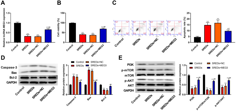
Rat model of TLE was produced by intraperitoneal injection of lithium chloride and pilocarpine. Rat hippocampal neuronal model of SREDs was established by Mg-free treatment. MEG3 was overexpressed by transfection of AAV-MEG3 in TLE and SREDs model. The expression of MEG3, interleukin-1β (IL-1β), interleukin-6 (IL-6) and recombinant human tumor necrosis factor-alpha (TNF-α) was detected by reverse transcription-quantitative polymerase chain reaction (RT-qPCR). Malondialdehyde (MDA) content and superoxide dismutase (SOD) activity were detected by corresponding kit. The apoptosis of hippocampal neurons was detected by terminal deoxynucleotidyl transferase transfer‑mediated dUTP nick end‑labeling (TUNEL) assay and flow cytometry. The expression of proteins related to apoptosis (Caspase-3, Bax, and Bcl-2) and the PI3K/AKT/mTOR pathway was detected by Western blot.
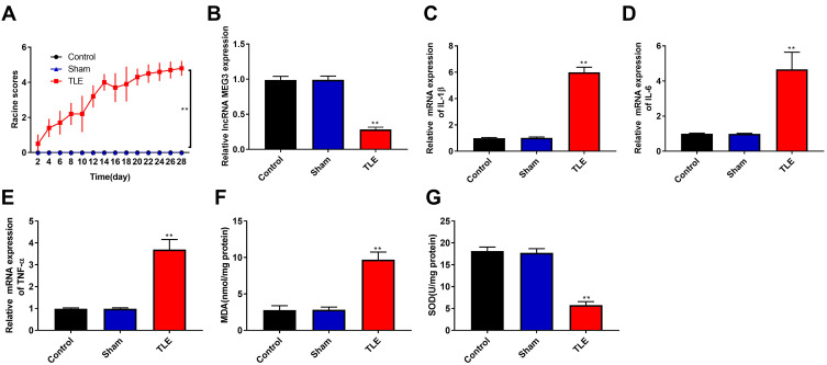
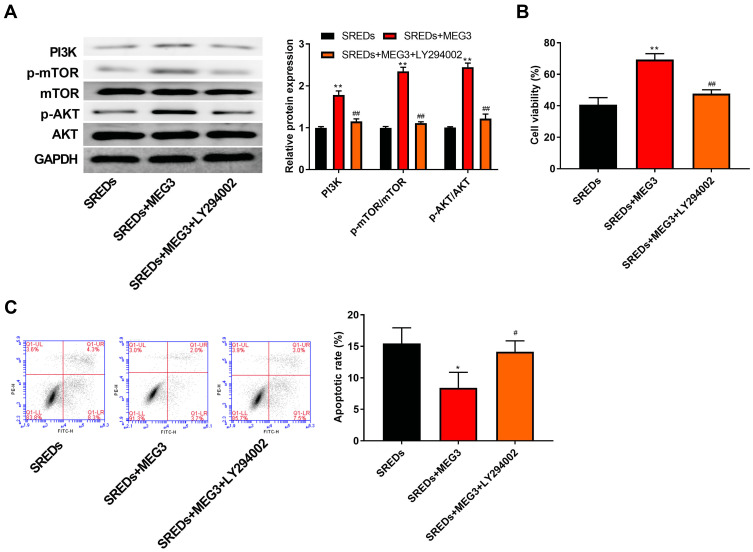
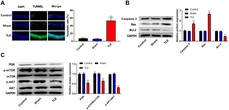
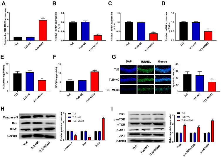
MEG3 expression was downregulated in SREDs and rats with TLE. Overexpression of MEG3 reduced the expression of IL-1β, IL-6, and TNF-α, MDA content, apoptosis rate of hippocampal neuron, increased SOD activity, and inhibited the PI3K/AKT/mTOR pathway in rats with TLE. In addition, overexpression of MEG3 enhanced cell viability and inhibited apoptosis through the activation of the PI3K/AKT/mTOR pathway in SREDs.
MEG3 reduced proinflammatory cytokines, oxidative stress, and apoptosis rate of hippocampal neuron and enhanced cell viability through the activation of the PI3K/AKT/mTOR pathway in SREDs and rats with TLE. Our findings may contribute to find a new therapeutic target for the treatment of epilepsy.
颞叶癫痫(TLE)是一种常见的神经系统疾病,其特征为反复出现的自发性癫痫发作。探索癫痫发生机制一直被视为首要任务。本研究旨在探讨长链非编码RNA MEG3在自发性反复癫痫样放电(SREDs)及TLE大鼠中的作用。
通过腹腔注射氯化锂和匹罗卡品制备TLE大鼠模型。通过无镁处理建立SREDs大鼠海马神经元模型。在TLE和SREDs模型中,通过转染腺相关病毒-MEG3(AAV-MEG3)使MEG3过表达。采用逆转录-定量聚合酶链反应(RT-qPCR)检测MEG3、白细胞介素-1β(IL-1β)、白细胞介素-6(IL-6)和重组人肿瘤坏死因子-α(TNF-α)的表达。使用相应试剂盒检测丙二醛(MDA)含量和超氧化物歧化酶(SOD)活性。通过末端脱氧核苷酸转移酶介导的dUTP缺口末端标记(TUNEL)法和流式细胞术检测海马神经元的凋亡情况。采用蛋白质印迹法检测与凋亡相关的蛋白(半胱天冬酶-3、Bax和Bcl-2)以及PI3K/AKT/mTOR信号通路的表达。
在SREDs及TLE大鼠中MEG3表达下调。MEG3过表达降低了TLE大鼠中IL-1β、IL-6和TNF-α的表达、MDA含量、海马神经元凋亡率,增加了SOD活性,并抑制了PI3K/AKT/mTOR信号通路。此外,MEG3过表达通过激活SREDs中的PI3K/AKT/mTOR信号通路增强了细胞活力并抑制了凋亡。
MEG3通过激活SREDs及TLE大鼠中的PI3K/AKT/mTOR信号通路,降低了促炎细胞因子、氧化应激和海马神经元凋亡率,并增强了细胞活力。我们的研究结果可能有助于找到治疗癫痫的新治疗靶点。