Department of Biological Sciences, Graduate School of Science, The University of Tokyo, Bunkyo, Tokyo, 113-0033, Japan.
Department of Life Science and Applied Chemistry, Nagoya Institute of Technology, Showa-Ku, Nagoya, 466-8555, Japan.
Nat Commun. 2020 Nov 5;11(1):5605. doi: 10.1038/s41467-020-19376-7.
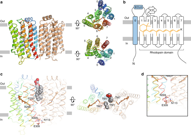
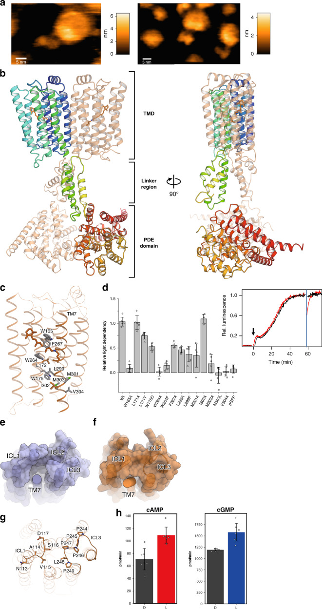
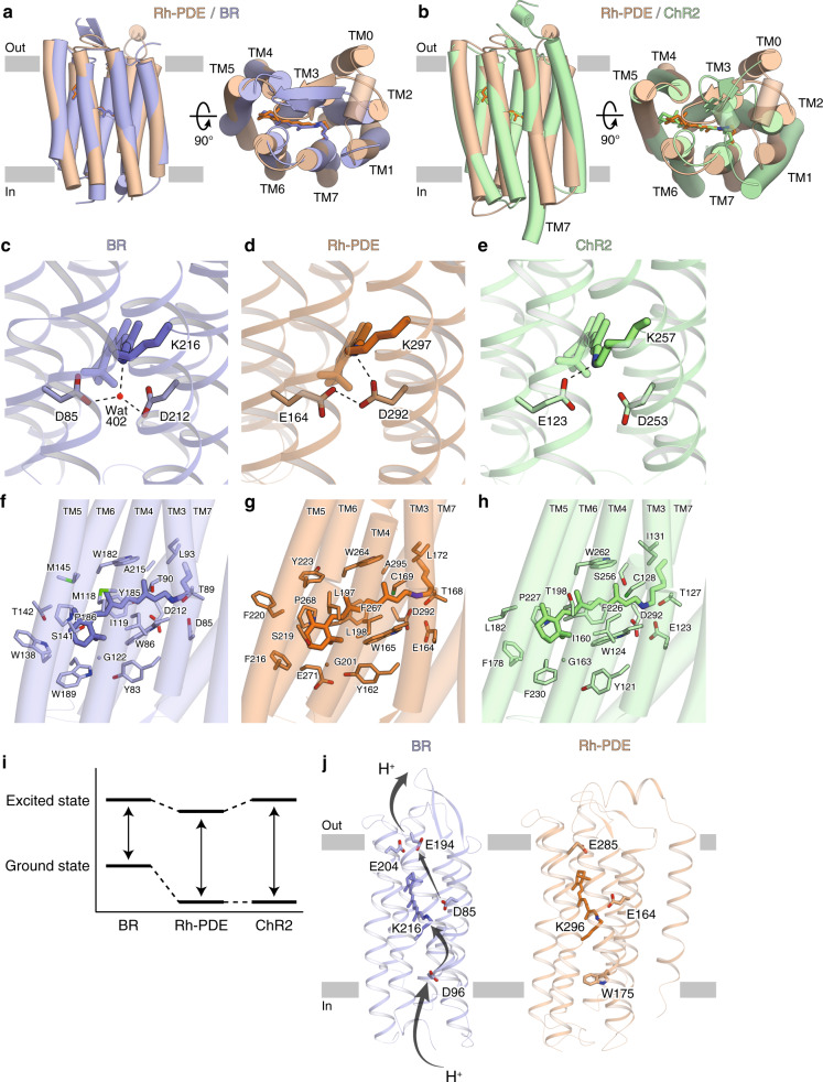
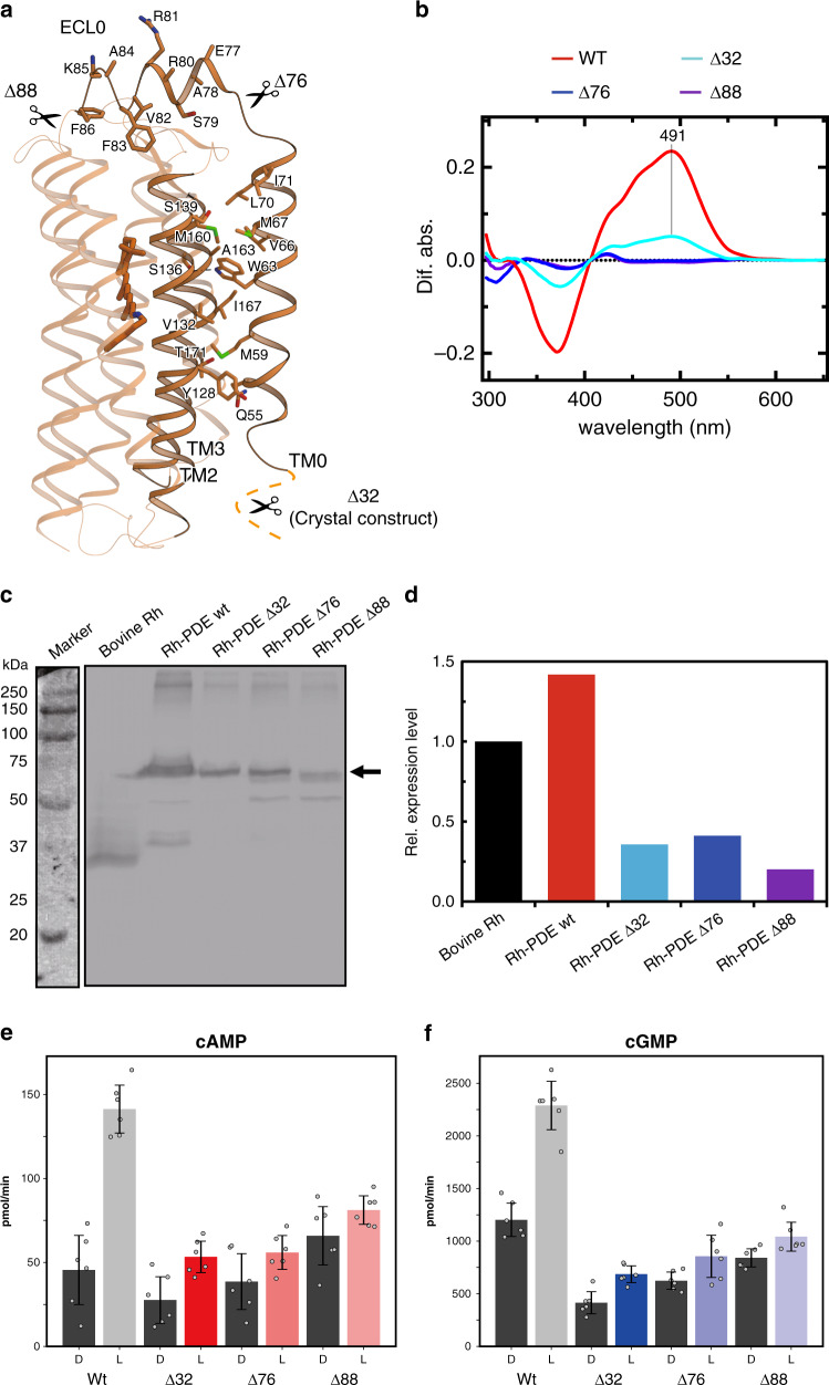
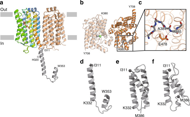
Rhodopsin phosphodiesterase (Rh-PDE) is an enzyme rhodopsin belonging to a recently discovered class of microbial rhodopsins with light-dependent enzymatic activity. Rh-PDE consists of the N-terminal rhodopsin domain and C-terminal phosphodiesterase (PDE) domain, connected by 76-residue linker, and hydrolyzes both cAMP and cGMP in a light-dependent manner. Thus, Rh-PDE has potential for the optogenetic manipulation of cyclic nucleotide concentrations, as a complementary tool to rhodopsin guanylyl cyclase and photosensitive adenylyl cyclase. Here we present structural and functional analyses of the Rh-PDE derived from Salpingoeca rosetta. The crystal structure of the rhodopsin domain at 2.6 Å resolution revealed a new topology of rhodopsins, with 8 TMs including the N-terminal extra TM, TM0. Mutational analyses demonstrated that TM0 plays a crucial role in the enzymatic photoactivity. We further solved the crystal structures of the rhodopsin domain (3.5 Å) and PDE domain (2.1 Å) with their connecting linkers, which showed a rough sketch of the full-length Rh-PDE. Integrating these structures, we proposed a model of full-length Rh-PDE, based on the HS-AFM observations and computational modeling of the linker region. These findings provide insight into the photoactivation mechanisms of other 8-TM enzyme rhodopsins and expand the definition of rhodopsins.
视紫红质磷酸二酯酶(Rh-PDE)是一种酶视紫红质,属于最近发现的具有光依赖性酶活性的微生物视紫红质类。Rh-PDE 由 N 端视紫红质结构域和 C 端磷酸二酯酶(PDE)结构域组成,通过 76 个残基的连接子连接,以光依赖性方式水解 cAMP 和 cGMP。因此,Rh-PDE 有望成为光遗传学操纵环核苷酸浓度的工具,作为视紫红质鸟苷酸环化酶和光敏腺苷酸环化酶的补充工具。在这里,我们介绍了来自罗塞塔氏管腔菌的 Rh-PDE 的结构和功能分析。2.6 Å 分辨率的视紫红质结构域的晶体结构揭示了一种新的视紫红质拓扑结构,包含 8 个跨膜结构域,包括 N 端额外的 TM0 结构域。突变分析表明 TM0 在酶的光活性中起着至关重要的作用。我们进一步解析了与连接子相连的视紫红质结构域(3.5 Å)和 PDE 结构域(2.1 Å)的晶体结构,展示了全长 Rh-PDE 的大致轮廓。综合这些结构,我们提出了全长 Rh-PDE 的模型,该模型基于 HS-AFM 观察和连接区的计算建模。这些发现为其他 8-TM 酶视紫红质的光激活机制提供了深入了解,并扩展了视紫红质的定义。