Department of Neurology, Neurovascular Research Group, Goethe University Hospital Frankfurt, Schleusenweg 2-16, 60528, Frankfurt, Germany.
Institute of Neuroradiology, Goethe University, Frankfurt, Germany.
Dysphagia. 2021 Oct;36(5):882-890. doi: 10.1007/s00455-020-10204-0. Epub 2020 Nov 6.
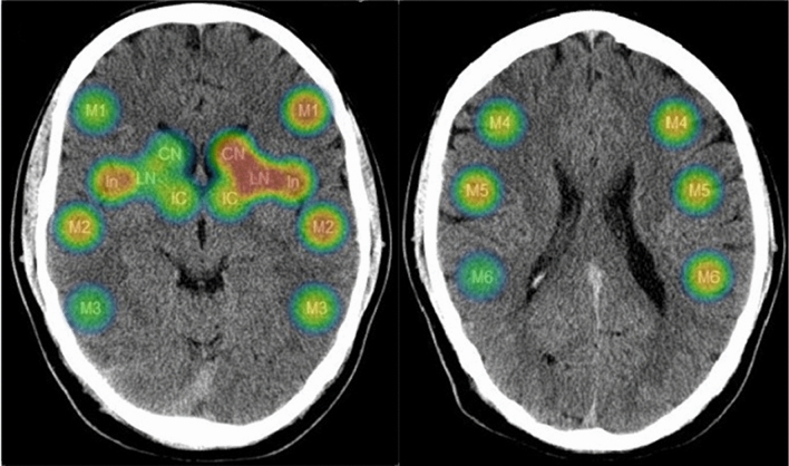
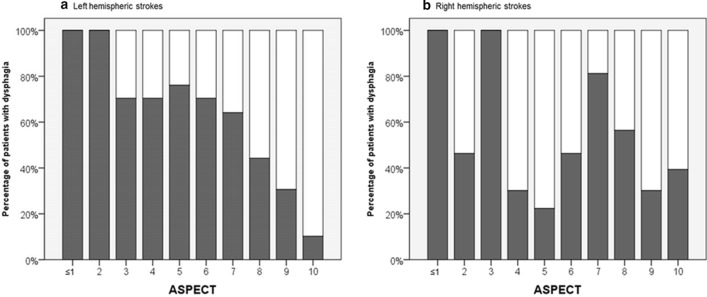
Dysphagia is common in patients with middle cerebral artery (MCA) infarctions and associated with malnutrition, pneumonia, and mortality. Besides bedside screening tools, brain imaging findings may help to timely identify patients with swallowing disorders. We investigated whether the Alberta stroke program early CT score (ASPECTS) allows for the correlation of distinct ischemic lesion patterns with dysphagia. We prospectively examined 113 consecutive patients with acute MCA infarctions. Fiberoptic endoscopic evaluation of swallowing (FEES) was performed within 24 h after admission for validation of dysphagia. Brain imaging (CT or MRI) was rated for ischemic changes according to the ASPECT score. 62 patients (54.9%) had FEES-proven dysphagia. In left hemispheric strokes, the strongest associations between the ASPECTS sectors and dysphagia were found for the lentiform nucleus (odds ratio 0.113 [CI 0.028-0.433; p = 0.001), the insula (0.275 [0.102-0.742]; p = 0.011), and the frontal operculum (0.280 [CI 0.094-0.834]; p = 0.022). A combination of two or even all three of these sectors together increased relative dysphagia frequency up to 100%. For right hemispheric strokes, only non-significant associations were found which were strongest for the insula region. The distribution of early ischemic changes in the MCA territory according to ASPECTS may be used as risk indicator of neurogenic dysphagia in MCA infarction, particularly when the left hemisphere is affected. However, due to the exploratory nature of this research, external validation studies of these findings are warranted in future.
大脑中动脉(MCA)梗死患者常伴有吞咽困难,可导致营养不良、肺炎和死亡。除了床边筛查工具外,脑影像学表现也可能有助于及时识别吞咽障碍患者。我们研究了 Alberta 卒中项目早期 CT 评分(ASPECTS)是否可以将不同的缺血性病变模式与吞咽困难相关联。我们前瞻性地检查了 113 例连续的急性 MCA 梗死患者。在入院后 24 小时内进行纤维光学内镜吞咽评估(FEES)以验证吞咽困难。根据 ASPECT 评分对脑影像学(CT 或 MRI)进行缺血性改变的评分。62 例患者(54.9%)经 FEES 证实存在吞咽困难。在左侧半球卒中患者中,ASPECTS 各区域与吞咽困难之间存在最强的相关性,与豆状核(比值比 0.113 [CI 0.028-0.433;p=0.001)、岛叶(0.275 [0.102-0.742];p=0.011)和额盖(0.280 [CI 0.094-0.834];p=0.022)关系最密切。即使是两个或三个区域的组合,也会使相对吞咽困难的频率增加到 100%。对于右侧半球卒中患者,仅发现了非显著相关性,而这些相关性在岛叶区域最为明显。根据 ASPECTS 对 MCA 区域的早期缺血性改变进行分布,可能成为 MCA 梗死神经源性吞咽困难的风险指标,尤其是当左侧半球受累时。然而,由于本研究的探索性质,需要在未来进行这些发现的外部验证研究。