• 文献检索
  • 文档翻译
  • 深度研究
  • 学术资讯
  • Suppr Zotero 插件Zotero 插件
  • 邀请有礼
  • 套餐&价格
  • 历史记录
应用&插件
Suppr Zotero 插件Zotero 插件浏览器插件Mac 客户端Windows 客户端微信小程序
定价
高级版会员购买积分包购买API积分包
服务
文献检索文档翻译深度研究API 文档MCP 服务
关于我们
关于 Suppr公司介绍联系我们用户协议隐私条款
关注我们

Suppr 超能文献

核心技术专利:CN118964589B侵权必究
粤ICP备2023148730 号-1Suppr @ 2026

文献检索

告别复杂PubMed语法,用中文像聊天一样搜索,搜遍4000万医学文献。AI智能推荐,让科研检索更轻松。

立即免费搜索

文件翻译

保留排版,准确专业,支持PDF/Word/PPT等文件格式,支持 12+语言互译。

免费翻译文档

深度研究

AI帮你快速写综述,25分钟生成高质量综述,智能提取关键信息,辅助科研写作。

立即免费体验

激光捕获显微切割 RNA 微测序揭示视神经挤压相关早期视网膜神经节细胞层 mRNA 变化。

Laser Capture Microdissection-Based RNA Microsequencing Reveals Optic Nerve Crush-Related Early mRNA Alterations in Retinal Ganglion Cell Layer.

机构信息

Department of Ophthalmology, Changhai Hospital, Second Military Medical University, Shanghai, China.

Shanghai Key Lab of Cell Engineering, Shanghai, China.

出版信息

Transl Vis Sci Technol. 2020 Oct 27;9(11):30. doi: 10.1167/tvst.9.11.30. eCollection 2020 Oct.

DOI:10.1167/tvst.9.11.30
PMID:33173609
原文链接:https://pmc.ncbi.nlm.nih.gov/articles/PMC7594581/
Abstract

PURPOSE

To establish a method of laser capture microdissection (LCM) and RNA microsequencing for exploring optic nerve crush (ONC)-related early mRNA alterations in retinal ganglion cell (RGC) layer.

METHODS

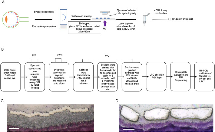
An LCM protocol was developed using retinal tissue sections to obtain high-quality RNA for microsequencing. Cells in the RGC layer were collected by laser pressure catapulting (LPC) using a PALM Zeiss UV LCM system. The effect of section thickness and slide type on tissue capture success and RNA yield and the integrity after LCM were evaluated. The optimal LCM protocol was used to explore ONC-related early mRNA alterations in the RGC layer. Candidate genes were validated by real-time polymerase chain reaction of the RGC layer tissue dissected by "cut and LPC" using the same LCM system.

RESULTS

We successfully established an optimal LCM protocol using 30-µm-thick retinal tissue sections mounted on glass slides and laser pressure catapulting (LPC) to collect cells in the RGC layer and to obtain high-quality RNA for microsequencing. On the basis of our protocol, we identified 8744 differentially expressed genes that were involved in ONC-related early mRNA alterations in the RGC layer. Candidate genes included Atf3, Lgals3, LOC102551701, Plaur, Tmem140, and Maml1.

CONCLUSIONS

The LCM-based single-cell RNA sequencing allowed a new sight into the early mRNA changes of RGCs highlighting new molecules associated to ONC.

TRANSLATIONAL RELEVANCE

This technique will be helpful for more accurate transcriptome analysis of clinical pathological samples of ophthalmology and provide important reference for the discovery of new pathological diagnosis indicators and drug development targets.

摘要

目的

建立激光捕获显微切割(LCM)和 RNA 微测序方法,以探索视神经挤压(ONC)相关的早期 RGC 层 mRNA 变化。

方法

使用视网膜组织切片开发了一种 LCM 方案,以获得用于微测序的高质量 RNA。使用 PALM Zeiss UV LCM 系统通过激光压力弹射(LPC)收集 RGC 层中的细胞。评估切片厚度和载玻片类型对组织捕获成功率、RNA 产量和 LCM 后 RNA 完整性的影响。使用最佳的 LCM 方案探索 RGC 层中与 ONC 相关的早期 mRNA 变化。使用相同的 LCM 系统对“切割和 LPC”分离的 RGC 层组织进行实时聚合酶链反应,验证候选基因。

结果

我们成功地使用 30-µm 厚的载玻片上的视网膜组织切片和激光压力弹射(LPC)建立了最佳的 LCM 方案,以收集 RGC 层中的细胞并获得用于微测序的高质量 RNA。在此方案的基础上,我们鉴定了 8744 个差异表达基因,这些基因参与了 RGC 层中与 ONC 相关的早期 mRNA 变化。候选基因包括 Atf3、Lgals3、LOC102551701、Plaur、Tmem140 和 Maml1。

结论

基于 LCM 的单细胞 RNA 测序为 RGC 早期 mRNA 变化提供了新的视角,突出了与 ONC 相关的新分子。

翻译的译文是

目的:建立激光捕获显微切割(LCM)和 RNA 微测序方法,以探索视神经挤压(ONC)相关的早期 RGC 层 mRNA 变化。

方法

使用视网膜组织切片开发了一种 LCM 方案,以获得用于微测序的高质量 RNA。使用 PALM Zeiss UV LCM 系统通过激光压力弹射(LPC)收集 RGC 层中的细胞。评估切片厚度和载玻片类型对组织捕获成功率、RNA 产量和 LCM 后 RNA 完整性的影响。使用最佳的 LCM 方案探索 RGC 层中与 ONC 相关的早期 mRNA 变化。使用相同的 LCM 系统对“切割和 LPC”分离的 RGC 层组织进行实时聚合酶链反应,验证候选基因。

结果

我们成功地使用 30-µm 厚的载玻片上的视网膜组织切片和激光压力弹射(LPC)建立了最佳的 LCM 方案,以收集 RGC 层中的细胞并获得用于微测序的高质量 RNA。在此方案的基础上,我们鉴定了 8744 个差异表达基因,这些基因参与了 RGC 层中与 ONC 相关的早期 mRNA 变化。候选基因包括 Atf3、Lgals3、LOC102551701、Plaur、Tmem140 和 Maml1。

结论

基于 LCM 的单细胞 RNA 测序为 RGC 早期 mRNA 变化提供了新的视角,突出了与 ONC 相关的新分子。

https://cdn.ncbi.nlm.nih.gov/pmc/blobs/20a3/7594581/12d933678c55/tvst-9-11-30-f004.jpg
https://cdn.ncbi.nlm.nih.gov/pmc/blobs/20a3/7594581/05d26a0706ef/tvst-9-11-30-f001.jpg
https://cdn.ncbi.nlm.nih.gov/pmc/blobs/20a3/7594581/5936394028c8/tvst-9-11-30-f002.jpg
https://cdn.ncbi.nlm.nih.gov/pmc/blobs/20a3/7594581/0a9e107719a4/tvst-9-11-30-f003.jpg
https://cdn.ncbi.nlm.nih.gov/pmc/blobs/20a3/7594581/12d933678c55/tvst-9-11-30-f004.jpg

相似文献

1
Laser Capture Microdissection-Based RNA Microsequencing Reveals Optic Nerve Crush-Related Early mRNA Alterations in Retinal Ganglion Cell Layer.激光捕获显微切割 RNA 微测序揭示视神经挤压相关早期视网膜神经节细胞层 mRNA 变化。
Transl Vis Sci Technol. 2020 Oct 27;9(11):30. doi: 10.1167/tvst.9.11.30. eCollection 2020 Oct.
2
LCM-Seq for Retinal Cell Layer-Specific Responses During Optic Nerve Regeneration.LCM-Seq 用于视神经再生过程中视网膜细胞层特异性反应。
Methods Mol Biol. 2023;2636:311-321. doi: 10.1007/978-1-0716-3012-9_17.
3
Early Gene Expression Profile in Retinal Ganglion Cell Layer After Optic Nerve Crush in Mice.视神经夹伤后小鼠视网膜神经节细胞层早期基因表达谱。
Invest Ophthalmol Vis Sci. 2018 Jan 1;59(1):370-380. doi: 10.1167/iovs.17-22438.
4
Activation of the BMP4/Smad1 Pathway Promotes Retinal Ganglion Cell Survival and Axon Regeneration.BMP4/Smad1 通路的激活促进视网膜神经节细胞存活和轴突再生。
Invest Ophthalmol Vis Sci. 2019 Apr 1;60(5):1748-1759. doi: 10.1167/iovs.18-26449.
5
Programmed cell death-1 is expressed in large retinal ganglion cells and is upregulated after optic nerve crush.程序性细胞死亡蛋白1在大型视网膜神经节细胞中表达,在视神经挤压后上调。
Exp Eye Res. 2015 Nov;140:1-9. doi: 10.1016/j.exer.2015.08.008. Epub 2015 Aug 13.
6
Laser capture microdissection protocol for gene expression analysis in the brain.用于大脑基因表达分析的激光捕获显微切割方案。
Histochem Cell Biol. 2017 Sep;148(3):299-311. doi: 10.1007/s00418-017-1585-1. Epub 2017 May 31.
7
Neuroprotective effects of inhibitors of Acid-Sensing ion channels (ASICs) in optic nerve crush model in rodents.酸敏感离子通道(ASICs)抑制剂在啮齿动物视神经挤压模型中的神经保护作用。
Curr Eye Res. 2018 Jan;43(1):84-95. doi: 10.1080/02713683.2017.1383442. Epub 2017 Nov 7.
8
Effects of a conventional photocoagulator and a 3-ns pulse laser on preconditioning responses and retinal ganglion cell survival after optic nerve crush.传统光凝器和3纳秒脉冲激光对视神经挤压后预处理反应及视网膜神经节细胞存活的影响。
Exp Eye Res. 2014 Oct;127:77-90. doi: 10.1016/j.exer.2014.07.008. Epub 2014 Jul 21.
9
Molecular, anatomical and functional changes in the retinal ganglion cells after optic nerve crush in mice.小鼠视神经挤压后视网膜神经节细胞的分子、解剖学和功能变化
Doc Ophthalmol. 2015 Apr;130(2):149-56. doi: 10.1007/s10633-014-9478-2. Epub 2015 Jan 6.
10
Activating Transcription Factor 3 (ATF3) Protects Retinal Ganglion Cells and Promotes Functional Preservation After Optic Nerve Crush.激活转录因子 3(ATF3)在视神经挤压后保护视网膜神经节细胞并促进功能保留。
Invest Ophthalmol Vis Sci. 2020 Feb 7;61(2):31. doi: 10.1167/iovs.61.2.31.

引用本文的文献

1
Evaluation of Immunohistochemical Biomarkers in Diabetic Wistar Rats with Periodontal Disease.糖尿病性牙周病Wistar大鼠免疫组化生物标志物的评估
J Pers Med. 2024 May 15;14(5):527. doi: 10.3390/jpm14050527.

本文引用的文献

1
Activating Transcription Factor 3 (ATF3) Protects Retinal Ganglion Cells and Promotes Functional Preservation After Optic Nerve Crush.激活转录因子 3(ATF3)在视神经挤压后保护视网膜神经节细胞并促进功能保留。
Invest Ophthalmol Vis Sci. 2020 Feb 7;61(2):31. doi: 10.1167/iovs.61.2.31.
2
Single-Cell Profiles of Retinal Ganglion Cells Differing in Resilience to Injury Reveal Neuroprotective Genes.单细胞分析揭示了对损伤具有不同抵抗力的视网膜神经节细胞的特征,并发现了神经保护基因。
Neuron. 2019 Dec 18;104(6):1039-1055.e12. doi: 10.1016/j.neuron.2019.11.006. Epub 2019 Nov 26.
3
Galectin-3 is required for the microglia-mediated brain inflammation in a model of Huntington's disease.
半乳糖凝集素-3 是亨廷顿病模型中小胶质细胞介导的脑炎症所必需的。
Nat Commun. 2019 Aug 2;10(1):3473. doi: 10.1038/s41467-019-11441-0.
4
Neuroinflammation induced by the peptide amyloid-β (25-35) increase the presence of galectin-3 in astrocytes and microglia and impairs spatial memory.肽淀粉样-β(25-35)引起的神经炎症增加了星形胶质细胞和小胶质细胞中半乳糖凝集素-3 的存在,并损害了空间记忆。
Neuropeptides. 2019 Apr;74:11-23. doi: 10.1016/j.npep.2019.02.001. Epub 2019 Feb 14.
5
Molecular Classification and Comparative Taxonomics of Foveal and Peripheral Cells in Primate Retina.灵长类动物视网膜中央凹和周边细胞的分子分类和比较分类学。
Cell. 2019 Feb 21;176(5):1222-1237.e22. doi: 10.1016/j.cell.2019.01.004. Epub 2019 Jan 31.
6
Early Gene Expression Profile in Retinal Ganglion Cell Layer After Optic Nerve Crush in Mice.视神经夹伤后小鼠视网膜神经节细胞层早期基因表达谱。
Invest Ophthalmol Vis Sci. 2018 Jan 1;59(1):370-380. doi: 10.1167/iovs.17-22438.
7
Immuno-Laser-Capture Microdissection for the Isolation of Enriched Glial Populations from Frozen Post-Mortem Human Brain.免疫激光捕获显微切割技术用于从冷冻的人死后大脑中分离富集的神经胶质细胞群体。
Methods Mol Biol. 2018;1723:273-284. doi: 10.1007/978-1-4939-7558-7_16.
8
Cell Type-Specific Laser Capture Microdissection for Gene Expression Profiling in the Human Brain.用于人类大脑基因表达谱分析的细胞类型特异性激光捕获显微切割技术
Methods Mol Biol. 2018;1723:203-221. doi: 10.1007/978-1-4939-7558-7_11.
9
Laser capture microdissection protocol for gene expression analysis in the brain.用于大脑基因表达分析的激光捕获显微切割方案。
Histochem Cell Biol. 2017 Sep;148(3):299-311. doi: 10.1007/s00418-017-1585-1. Epub 2017 May 31.
10
Using Laser Capture Microdissection to Isolate Cortical Laminae in Nonhuman Primate Brain.利用激光捕获显微切割技术分离非人灵长类动物大脑中的皮质层。
Methods Mol Biol. 2017;1606:115-132. doi: 10.1007/978-1-4939-6990-6_8.