Department of Neurology, Neuroinnovation Program, Multiple Sclerosis and Neuroimmunology Imaging Program, Clinical Center for Multiple Sclerosis, UT Southwestern Medical Center, 5323 Harry Hines Blvd., Dallas, TX, 75390-8806, USA.
School of Medicine, UT Southwestern Medical Center, 6011 Harry Hines Blvd., Dallas, TX, 75235, USA.
Sci Rep. 2020 Nov 11;10(1):19560. doi: 10.1038/s41598-020-76420-8.
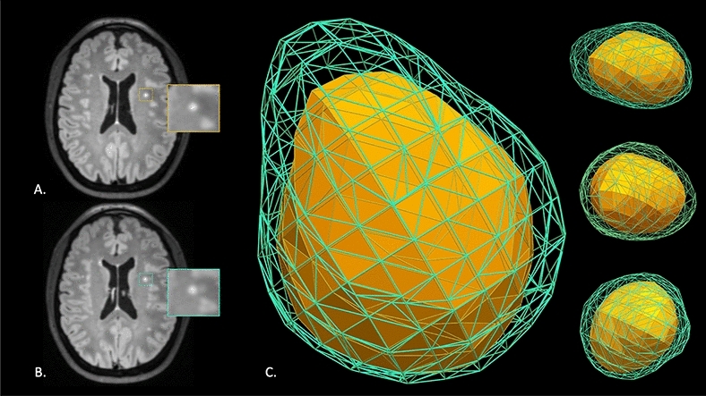
The accurate recognition of multiple sclerosis (MS) lesions is challenged by the high sensitivity and imperfect specificity of MRI. To examine whether longitudinal changes in volume, surface area, 3-dimensional (3D) displacement (i.e. change in lesion position), and 3D deformation (i.e. change in lesion shape) could inform on the origin of supratentorial brain lesions, we prospectively enrolled 23 patients with MS and 11 patients with small vessel disease (SVD) and performed standardized 3-T 3D brain MRI studies. Bayesian linear mixed effects regression models were constructed to evaluate associations between changes in lesion morphology and disease state. A total of 248 MS and 157 SVD lesions were studied. Individual MS lesions demonstrated significant decreases in volume < 3.75mm (p = 0.04), greater shifts in 3D displacement by 23.4% with increasing duration between MRI time points (p = 0.007), and greater transitions to a more non-spherical shape (p < 0.0001). If 62.2% of lesions within a given MRI study had a calculated theoretical radius > 2.49 based on deviation from a perfect 3D sphere, a 92.7% in-sample and 91.2% out-of-sample accuracy was identified for the diagnosis of MS. Longitudinal 3D shape evolution and displacement characteristics may improve lesion classification, adding to MRI techniques aimed at improving lesion specificity.
多发性硬化症(MS)病变的准确识别受到 MRI 高灵敏度和不完善特异性的挑战。为了研究体积、表面积、三维(3D)位移(即病变位置的变化)和 3D 变形(即病变形状的变化)的纵向变化是否可以提示幕上脑病变的起源,我们前瞻性地招募了 23 例 MS 患者和 11 例小血管疾病(SVD)患者,并进行了标准化的 3T 3D 脑部 MRI 研究。构建了贝叶斯线性混合效应回归模型来评估病变形态变化与疾病状态之间的关联。共研究了 248 个 MS 和 157 个 SVD 病变。个体 MS 病变的体积明显减小<3.75mm(p=0.04),3D 位移的变化更大,在 MRI 时间点之间的间隔增加 23.4%(p=0.007),向更非球形的形状转变更大(p<0.0001)。如果给定 MRI 研究中计算的理论半径>2.49 的病变比例为 62.2%,基于对完美 3D 球体的偏差,则 MS 的诊断准确率为 92.7%(样本内)和 91.2%(样本外)。3D 形状的纵向演变和位移特征可能会改善病变分类,这增加了旨在提高病变特异性的 MRI 技术。