• 文献检索
  • 文档翻译
  • 深度研究
  • 学术资讯
  • Suppr Zotero 插件Zotero 插件
  • 邀请有礼
  • 套餐&价格
  • 历史记录
应用&插件
Suppr Zotero 插件Zotero 插件浏览器插件Mac 客户端Windows 客户端微信小程序
定价
高级版会员购买积分包购买API积分包
服务
文献检索文档翻译深度研究API 文档MCP 服务
关于我们
关于 Suppr公司介绍联系我们用户协议隐私条款
关注我们

Suppr 超能文献

核心技术专利:CN118964589B侵权必究
粤ICP备2023148730 号-1Suppr @ 2026

文献检索

告别复杂PubMed语法,用中文像聊天一样搜索,搜遍4000万医学文献。AI智能推荐,让科研检索更轻松。

立即免费搜索

文件翻译

保留排版,准确专业,支持PDF/Word/PPT等文件格式,支持 12+语言互译。

免费翻译文档

深度研究

AI帮你快速写综述,25分钟生成高质量综述,智能提取关键信息,辅助科研写作。

立即免费体验

水通道蛋白 5 表达的小鼠肺上皮细胞的单细胞 RNA 测序分析鉴定 GPRC5A 为新型的 I 型细胞表面标志物。

Integrated Single-Cell RNA-Sequencing Analysis of Aquaporin 5-Expressing Mouse Lung Epithelial Cells Identifies GPRC5A as a Novel Validated Type I Cell Surface Marker.

机构信息

Hastings Center for Pulmonary Research and Division of Pulmonary, Critical Care and Sleep Medicine, Department of Medicine, Keck School of Medicine, University of Southern California, Los Angeles, CA 90033, USA.

Department of Respiratory Medicine, Graduate School of Medicine, The University of Tokyo, 7-3-1 Hongo, Bunkyo-ku, Tokyo 113-0033, Japan.

出版信息

Cells. 2020 Nov 11;9(11):2460. doi: 10.3390/cells9112460.

DOI:10.3390/cells9112460
PMID:33187367
原文链接:https://pmc.ncbi.nlm.nih.gov/articles/PMC7697677/
Abstract

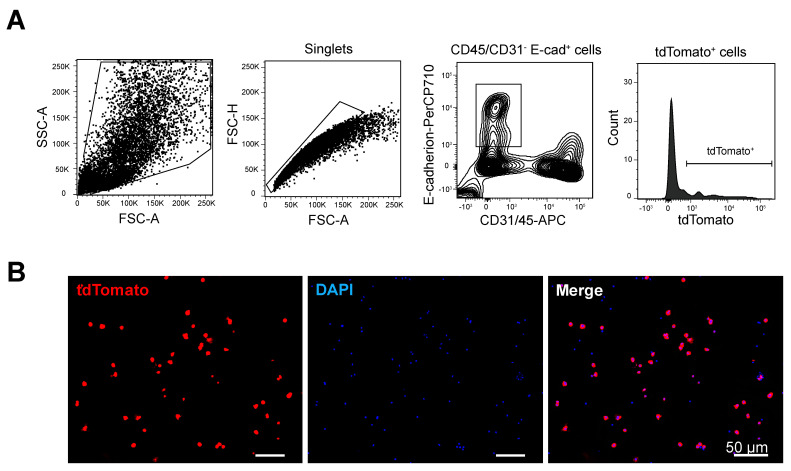
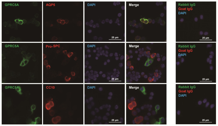
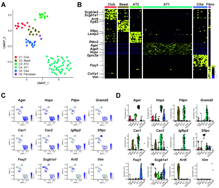
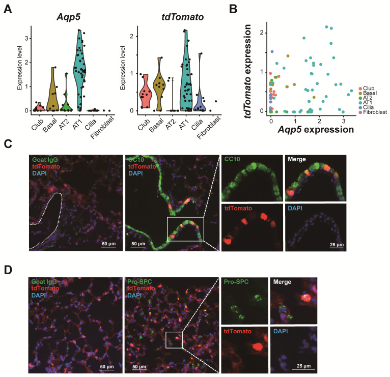
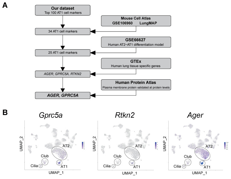
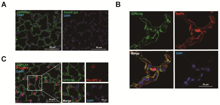
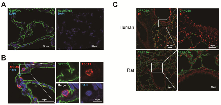
Molecular and functional characterization of alveolar epithelial type I (AT1) cells has been challenging due to difficulty in isolating sufficient numbers of viable cells. Here we performed single-cell RNA-sequencing (scRNA-seq) of tdTomato cells from lungs of AT1 cell-specific Aqp5-Cre-IRES-DsRed (ACID);R26tdTomato reporter mice. Following enzymatic digestion, CD31CD45E-cadherintdTomato cells were subjected to fluorescence-activated cell sorting (FACS) followed by scRNA-seq. Cell identity was confirmed by immunofluorescence using cell type-specific antibodies. After quality control, 92 cells were analyzed. Most cells expressed 'conventional' AT1 cell markers (, , , ), with heterogeneous expression within this population. The remaining cells expressed AT2, club, basal or ciliated cell markers. Integration with public datasets identified three robust AT1 cell- and lung-enriched genes, , and , that were conserved across species. GPRC5A co-localized with HOPX and was not expressed in AT2 or airway cells in mouse, rat and human lung. GPRC5A co-localized with AQP5 but not pro-SPC or CC10 in mouse lung epithelial cell cytospins. We enriched mouse AT1 cells to perform molecular phenotyping using scRNA-seq. Further characterization of putative AT1 cell-enriched genes revealed GPRC5A as a conserved AT1 cell surface marker that may be useful for AT1 cell isolation.

摘要

由于难以分离足够数量的活细胞,肺泡上皮细胞 I 型(AT1)的分子和功能特征一直具有挑战性。在这里,我们对 AT1 细胞特异性 Aqp5-Cre-IRES-DsRed(ACID);R26tdTomato 报告小鼠肺部的 tdTomato 细胞进行了单细胞 RNA 测序(scRNA-seq)。在进行酶消化后,使用 CD31CD45E-cadherintdTomato 细胞进行荧光激活细胞分选(FACS),然后进行 scRNA-seq。使用细胞类型特异性抗体通过免疫荧光确认细胞身份。经过质量控制后,分析了 92 个细胞。大多数细胞表达“传统”AT1 细胞标志物(,,,),在该群体中具有异质性表达。其余细胞表达 AT2、club、基底或纤毛细胞标志物。与公共数据集的整合确定了三个稳健的 AT1 细胞和肺富集基因,,和,这些基因在物种间是保守的。GPRC5A 与 HOPX 共定位,在小鼠、大鼠和人肺中的 AT2 或气道细胞中不表达。GPRC5A 与 AQP5 共定位,但与小鼠肺上皮细胞涂片中的 pro-SPC 或 CC10 不共定位。我们富集了小鼠 AT1 细胞,以使用 scRNA-seq 进行分子表型分析。对假定的 AT1 细胞富集基因的进一步表征揭示了 GPRC5A 作为一种保守的 AT1 细胞表面标志物,可能有助于 AT1 细胞的分离。

https://cdn.ncbi.nlm.nih.gov/pmc/blobs/fd7b/7697677/aed1b3dad396/cells-09-02460-g007.jpg
https://cdn.ncbi.nlm.nih.gov/pmc/blobs/fd7b/7697677/570c69cfc509/cells-09-02460-g001.jpg
https://cdn.ncbi.nlm.nih.gov/pmc/blobs/fd7b/7697677/086c0d8a5099/cells-09-02460-g002.jpg
https://cdn.ncbi.nlm.nih.gov/pmc/blobs/fd7b/7697677/6ed41928b31c/cells-09-02460-g003.jpg
https://cdn.ncbi.nlm.nih.gov/pmc/blobs/fd7b/7697677/aee15d24eb0c/cells-09-02460-g004a.jpg
https://cdn.ncbi.nlm.nih.gov/pmc/blobs/fd7b/7697677/5a7257e60c36/cells-09-02460-g005.jpg
https://cdn.ncbi.nlm.nih.gov/pmc/blobs/fd7b/7697677/3c5cf66d96bb/cells-09-02460-g006.jpg
https://cdn.ncbi.nlm.nih.gov/pmc/blobs/fd7b/7697677/aed1b3dad396/cells-09-02460-g007.jpg

相似文献

1
Integrated Single-Cell RNA-Sequencing Analysis of Aquaporin 5-Expressing Mouse Lung Epithelial Cells Identifies GPRC5A as a Novel Validated Type I Cell Surface Marker.水通道蛋白 5 表达的小鼠肺上皮细胞的单细胞 RNA 测序分析鉴定 GPRC5A 为新型的 I 型细胞表面标志物。
Cells. 2020 Nov 11;9(11):2460. doi: 10.3390/cells9112460.
2
Combinations of differentiation markers distinguish subpopulations of alveolar epithelial cells in adult lung.分化标志物的组合可区分成年肺中肺泡上皮细胞的亚群。
Am J Physiol Lung Cell Mol Physiol. 2016 Jan 15;310(2):L114-20. doi: 10.1152/ajplung.00337.2015. Epub 2015 Nov 6.
3
Directed expression of Cre in alveolar epithelial type 1 cells.在肺泡上皮细胞 1 型中定向表达 Cre。
Am J Respir Cell Mol Biol. 2010 Aug;43(2):173-8. doi: 10.1165/rcmb.2009-0226OC. Epub 2009 Sep 18.
4
Fraction of MHCII and EpCAM expression characterizes distal lung epithelial cells for alveolar type 2 cell isolation.MHCII 和 EpCAM 表达的分数特征化了用于分离肺泡 2 型细胞的远端肺上皮细胞。
Respir Res. 2017 Aug 7;18(1):150. doi: 10.1186/s12931-017-0635-5.
5
Cross-Species Transcriptome Profiling Identifies New Alveolar Epithelial Type I Cell-Specific Genes.跨物种转录组分析鉴定出新的肺泡I型上皮细胞特异性基因。
Am J Respir Cell Mol Biol. 2017 Mar;56(3):310-321. doi: 10.1165/rcmb.2016-0071OC.
6
Pulmonary alveolar type I cell population consists of two distinct subtypes that differ in cell fate.肺泡 I 型细胞群体由两个不同的亚型组成,它们在细胞命运上有所不同。
Proc Natl Acad Sci U S A. 2018 Mar 6;115(10):2407-2412. doi: 10.1073/pnas.1719474115. Epub 2018 Feb 20.
7
Deciphering the molecular regulatory of RAB32/GPRC5A axis in chronic obstructive pulmonary disease.解析 RAB32/GPRC5A 轴在慢性阻塞性肺疾病中的分子调控机制。
Respir Res. 2024 Mar 6;25(1):116. doi: 10.1186/s12931-024-02724-2.
8
Single-cell transcriptome profiling reveals the mechanism of abnormal proliferation of epithelial cells in congenital cystic adenomatoid malformation.单细胞转录组谱分析揭示先天性囊性腺瘤样畸形中上皮细胞异常增殖的机制。
Exp Cell Res. 2020 Nov 15;396(2):112299. doi: 10.1016/j.yexcr.2020.112299. Epub 2020 Sep 23.
9
Impaired AT2 to AT1 cell transition in PM2.5-induced mouse model of chronic obstructive pulmonary disease.PM2.5 诱导的慢性阻塞性肺疾病小鼠模型中 AT2 向 AT1 细胞转化受损。
Respir Res. 2022 Mar 25;23(1):70. doi: 10.1186/s12931-022-01996-w.
10
Long-Term Engraftment Promotes Differentiation of Alveolar Epithelial Cells from Human Embryonic Stem Cell Derived Lung Organoids.长期植入促进人胚胎干细胞衍生的肺类器官中的肺泡上皮细胞的分化。
Stem Cells Dev. 2018 Oct 1;27(19):1339-1349. doi: 10.1089/scd.2018.0042. Epub 2018 Aug 21.

引用本文的文献

1
Direct reprogramming of mouse fibroblasts into self-renewable alveolar epithelial-like cells.将小鼠成纤维细胞直接重编程为可自我更新的肺泡上皮样细胞。
NPJ Regen Med. 2025 Jun 23;10(1):30. doi: 10.1038/s41536-025-00411-4.
2
Single-Cell Transcriptomics Reveals Stem Cell-Derived Exosomes Attenuate Inflammatory Gene Expression in Pulmonary Oxygen Toxicity.单细胞转录组学揭示干细胞衍生外泌体减轻肺氧中毒中的炎症基因表达。
Int J Mol Sci. 2025 May 7;26(9):4462. doi: 10.3390/ijms26094462.
3
Biomaterial-based 3D human lung models replicate pathological characteristics of early pulmonary fibrosis.

本文引用的文献

1
Single-Nucleus RNA-Sequencing Profiling of Mouse Lung. Reduced Dissociation Bias and Improved Rare Cell-Type Detection Compared with Single-Cell RNA Sequencing.单细胞 RNA 测序分析小鼠肺脏。与单细胞 RNA 测序相比,降低了解离偏倚,提高了稀有细胞类型的检测能力。
Am J Respir Cell Mol Biol. 2020 Dec;63(6):739-747. doi: 10.1165/rcmb.2020-0095MA.
2
Single-cell connectomic analysis of adult mammalian lungs.成年哺乳动物肺部的单细胞连接组学分析。
Sci Adv. 2019 Dec 4;5(12):eaaw3851. doi: 10.1126/sciadv.aaw3851. eCollection 2019 Dec.
3
Rare Pulmonary Neuroendocrine Cells Are Stem Cells Regulated by Rb, p53, and Notch.
基于生物材料的3D人体肺部模型可复制早期肺纤维化的病理特征。
bioRxiv. 2025 Feb 17:2025.02.12.637970. doi: 10.1101/2025.02.12.637970.
4
From Epithelium to Therapy: Transitional Cells in Lung Fibrosis.从上皮细胞到治疗:肺纤维化中的过渡细胞
Am J Respir Cell Mol Biol. 2025 May;72(5):472-483. doi: 10.1165/rcmb.2024-0372TR.
5
Integrated multiomic analysis identifies TRIP13 as a mediator of alveolar epithelial type II cell dysfunction in idiopathic pulmonary fibrosis.综合多组学分析确定TRIP13是特发性肺纤维化中II型肺泡上皮细胞功能障碍的介质。
Biochim Biophys Acta Mol Basis Dis. 2025 Mar;1871(3):167572. doi: 10.1016/j.bbadis.2024.167572. Epub 2024 Nov 13.
6
Single-cell transcriptome analysis of the mouse lungs during the injury and recovery periods after lipopolysaccharide administration.脂多糖给药后小鼠肺损伤和恢复期的单细胞转录组分析。
Inflamm Res. 2024 Dec;73(12):2087-2107. doi: 10.1007/s00011-024-01951-z. Epub 2024 Oct 8.
7
FixNCut: A Practical Guide to Sample Preservation by Reversible Fixation for Single Cell Assays.《固定与切割:单细胞分析中通过可逆固定进行样本保存实用指南》
Bio Protoc. 2024 Sep 5;14(17):e5063. doi: 10.21769/BioProtoc.5063.
8
Different localization of P2X4 and P2X7 receptors in native mouse lung - lack of evidence for a direct P2X4-P2X7 receptor interaction.P2X4 和 P2X7 受体在天然小鼠肺中的不同定位 - 缺乏直接 P2X4-P2X7 受体相互作用的证据。
Front Immunol. 2024 Jun 17;15:1425938. doi: 10.3389/fimmu.2024.1425938. eCollection 2024.
9
Epigenetic modifications in the development of bronchopulmonary dysplasia: a review.支气管肺发育不良的发生发展中的表观遗传修饰:综述。
Pediatr Res. 2024 Aug;96(3):632-642. doi: 10.1038/s41390-024-03167-7. Epub 2024 Apr 3.
10
Single-cell transcriptomic analysis of radiation-induced lung injury in rat.大鼠放射性肺损伤的单细胞转录组分析。
Biomol Biomed. 2024 Sep 6;24(5):1331-1349. doi: 10.17305/bb.2024.10357.
罕见的肺神经内分泌细胞是由 Rb、p53 和 Notch 调控的干细胞。
Cell. 2019 Oct 3;179(2):403-416.e23. doi: 10.1016/j.cell.2019.09.010.
4
Single cell RNA sequencing identifies TGFβ as a key regenerative cue following LPS-induced lung injury.单细胞 RNA 测序鉴定 TGFβ 为 LPS 诱导的肺损伤后关键的再生信号。
JCI Insight. 2019 Mar 26;5(8):123637. doi: 10.1172/jci.insight.123637.
5
Single-Cell Transcriptomic Analysis of Human Lung Provides Insights into the Pathobiology of Pulmonary Fibrosis.单细胞转录组分析人类肺部组织为肺纤维化的病理生物学提供新见解。
Am J Respir Crit Care Med. 2019 Jun 15;199(12):1517-1536. doi: 10.1164/rccm.201712-2410OC.
6
A single-cell atlas of the airway epithelium reveals the CFTR-rich pulmonary ionocyte.气道上皮细胞的单细胞图谱揭示了富含 CFTR 的肺离子细胞。
Nature. 2018 Aug;560(7718):377-381. doi: 10.1038/s41586-018-0394-6. Epub 2018 Aug 1.
7
Ager-CreER: A New Genetic Tool for Studying Lung Alveolar Development, Homeostasis, and Repair.Ager-CreER:一种研究肺肺泡发育、稳态和修复的新遗传工具。
Am J Respir Cell Mol Biol. 2018 Dec;59(6):706-712. doi: 10.1165/rcmb.2018-0125OC.
8
Integrating single-cell transcriptomic data across different conditions, technologies, and species.整合不同条件、技术和物种的单细胞转录组数据。
Nat Biotechnol. 2018 Jun;36(5):411-420. doi: 10.1038/nbt.4096. Epub 2018 Apr 2.
9
Mapping the Mouse Cell Atlas by Microwell-Seq.通过微室测序绘制小鼠细胞图谱。
Cell. 2018 Feb 22;172(5):1091-1107.e17. doi: 10.1016/j.cell.2018.02.001.
10
Pulmonary alveolar type I cell population consists of two distinct subtypes that differ in cell fate.肺泡 I 型细胞群体由两个不同的亚型组成,它们在细胞命运上有所不同。
Proc Natl Acad Sci U S A. 2018 Mar 6;115(10):2407-2412. doi: 10.1073/pnas.1719474115. Epub 2018 Feb 20.