Gonzalez-Rivas Diego, Soultanis Konstantinos Marios, Garcia Alejandro, Yang Kaiyun, Qing Yue, Yie Linhua, Zhao Guangqiang, Chen Anning, Huang Yunchao, Li Guangjian, Jiang Gening
Thoracic Surgery Department, Tongji University Affiliated Shanghai Pulmonary Hospital, Shanghai, China.
Department of Thoracic Surgery and Minimally Invasive Thoracic Surgery Unit (UCTMI), Coruña University Hospital, Coruña, Spain.
J Thorac Dis. 2020 Oct;12(10):6198-6209. doi: 10.21037/jtd.2020.04.05.
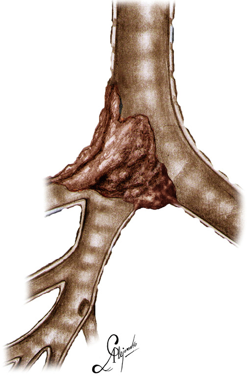
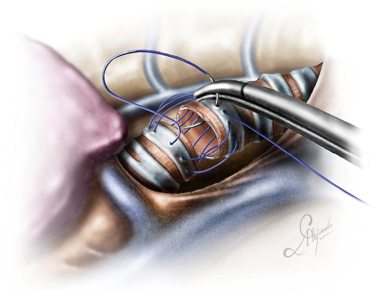
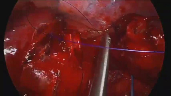
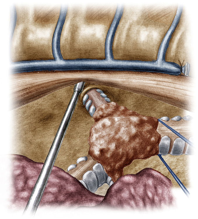
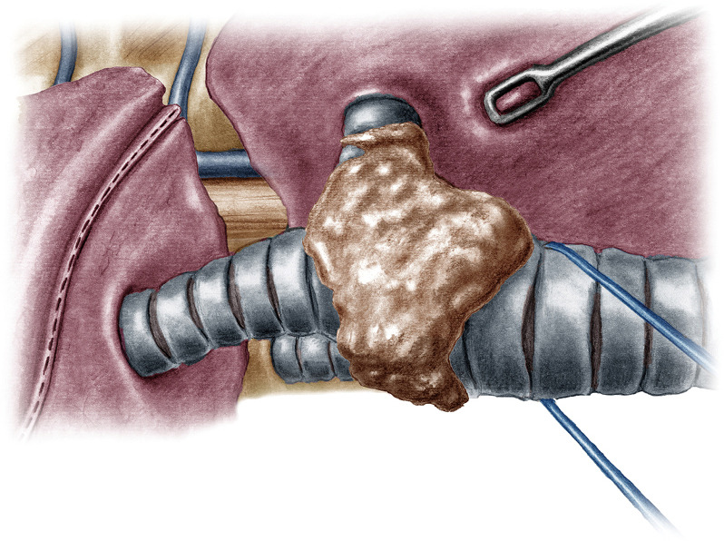
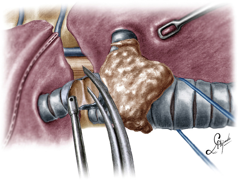
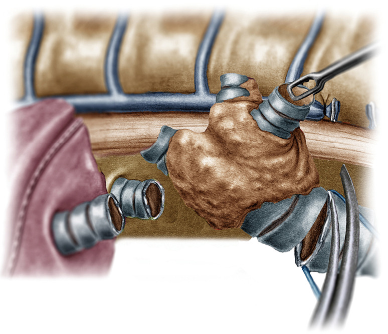
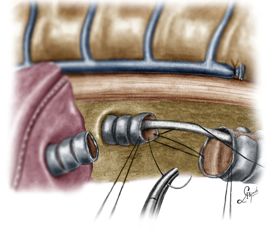
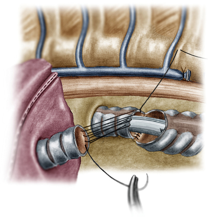
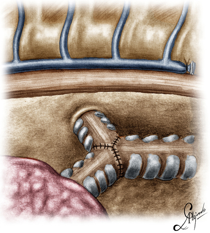
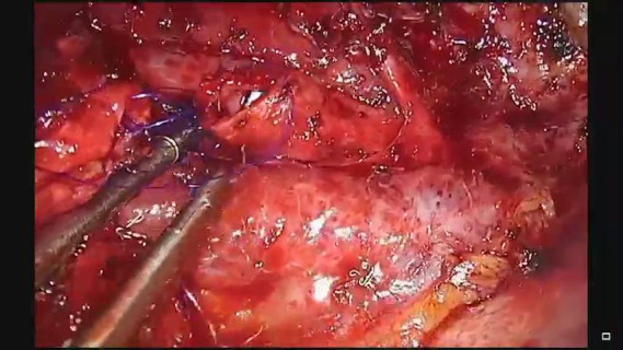
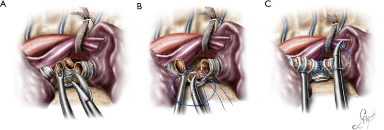
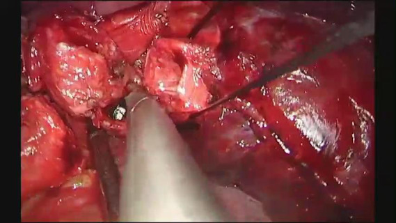
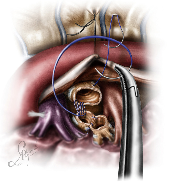
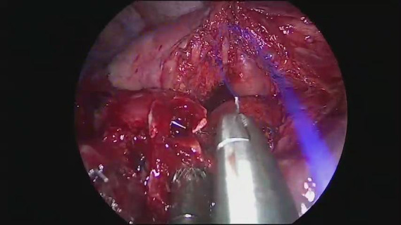
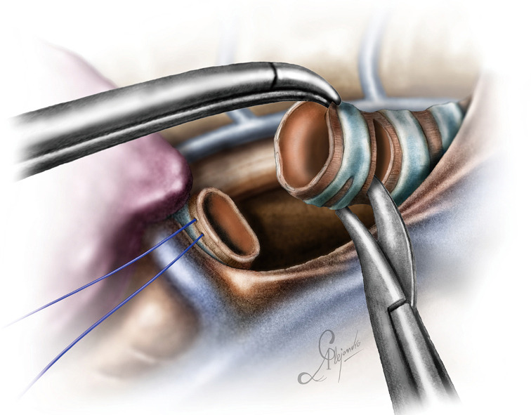
Pathology arising from the intrathoracic portion of the trachea (distal trachea), the carina and the main bronchi is usually neoplastic and is mainly treated with surgery. Resection of the intrathoracic portion of the trachea, the carina and the main bronchi for neoplastic lesions does not necessitate lung resection and is traditionally being conducted via open surgery. Video-assisted thoracic surgery (VATS) is witnessing an exponential growth and is the treatment of choice for early-stage non-small cell lung cancer (NSCLC). The experience accumulated over the past two decades along with the introduction of reliable and ergonomic technology, has led to the expansion of its indications. In this article we provide a detailed description of lung sparing distal tracheal, carinal and main bronchi resection for primary neoplasms of the airway, without involvement of the lung, with the uniportal video-assisted technique. The chest is entered through the fourth intercostal space, mid-axillary line. Dissection of the paratracheal space anteriorly, the tracheoesophageal groove posteriorly and the subcarinal space and division of the azygos arch are essential to mobilize the distal trachea and carina. Lateral dissection should be avoided beyond the points of division of the airway, as it may hinder the blood supply to the anastomosis. Any tension to the anastomosis should be relieved by release maneuvers. Ventilation is achieved through an endobronchial catheter, inserted into the left main bronchus through which a high-frequency jet ventilation catheter can be also inserted through it. The rationale of applying a minimally invasive technique for the conduction of tracheal and carinal resections, is to exploit its advantages, namely less pain, earlier mobilization and lower morbidity. Uniportal video-assisted resections of the distal trachea, carina and the main bronchi, are safe when conducted by experienced surgical and anesthetic teams.
起源于气管胸段(远端气管)、隆突和主支气管的病变通常为肿瘤性病变,主要通过手术治疗。因肿瘤性病变切除气管胸段、隆突和主支气管并不一定需要切除肺组织,传统上是通过开放手术进行。电视辅助胸腔镜手术(VATS)正在迅速发展,是早期非小细胞肺癌(NSCLC)的首选治疗方法。过去二十年积累的经验以及可靠且符合人体工程学的技术的引入,导致其适应证不断扩大。在本文中,我们详细描述了采用单孔电视辅助技术,在不涉及肺组织的情况下,对气道原发性肿瘤进行保留肺组织的远端气管、隆突和主支气管切除术。通过腋中线第四肋间间隙进入胸腔。在前方解剖气管旁间隙、后方解剖气管食管沟以及隆突下间隙并切断奇静脉弓,对于游离远端气管和隆突至关重要。应避免在气道分支点以外进行外侧解剖,因为这可能会阻碍吻合口的血供。任何对吻合口的张力都应通过松解操作来缓解。通过插入左主支气管的支气管内导管实现通气,高频喷射通气导管也可通过该导管插入。采用微创技术进行气管和隆突切除的基本原理是利用其优势,即疼痛减轻、活动更早和发病率更低。由经验丰富的手术和麻醉团队进行单孔电视辅助远端气管、隆突和主支气管切除术是安全的。