Department of Radiology, Dong-A University Medical Center, Busan, Republic of Korea.
Department of Neurology, Dong-A University Medical Center, Busan, Republic of Korea.
PLoS One. 2020 Nov 19;15(11):e0240911. doi: 10.1371/journal.pone.0240911. eCollection 2020.
The treatment strategy is different for acute traumatic peripheral nerve injury and acute compressive neuropathy. This study aimed to compare magnetic resonance imaging (MRI) features of acute traumatic peripheral nerve injury and acute compressive neuropathy in a rat model.
Twenty female Sprague-Dawley rats were divided into two groups. In the crush injury group (n = 10), the unilateral sciatic nerve was crushed using forceps to represent acute traumatic peripheral nerve injury. In the compression injury group (n = 10), the unilateral sciatic nerve was ligated using silk to represent acute compressive neuropathy. The MRI of eight rats from each group were acquired on postoperative days 3 and 10. Fat-suppressed T2-weighted images were acquired. Changes in the injured nerve were divided into three grades. A Fisher's exact test was used to compare the changes in the nerves of the two groups. Histological staining and a western blot analysis were performed on one rat in each group on day 3. Neurofilament, myelin basic protein (MBP), and p75NTR staining were performed. Expression of neurofilament, MBP, p75NTR, and c-jun was evaluated by western blot analysis.
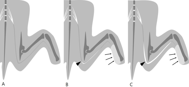
MR neurography revealed substantial nerve changes in the compression injury group compared with the crush injury group at two-time points (p = 0.001 on day 3, p = 0.026 on day 10). The histopathological analysis indicated the destruction of the axon and myelin, mainly at the injury site and the distal portion of the injury in the crush injury group. It was prominent in the proximal portion, the injury site, and the distal portion of the injury in the compression injury group. The degree of axonal and myelin destruction was more pronounced in the compression injury group than in the crush injury group.
MR neurography showed prominent and long-segmental changes associated with the injured nerve in acute compressive neuropathy compared with acute traumatic peripheral nerve injury.
急性创伤性周围神经损伤和急性压迫性神经病的治疗策略不同。本研究旨在比较大鼠模型中急性创伤性周围神经损伤和急性压迫性神经病的磁共振成像(MRI)特征。
将 20 只雌性 Sprague-Dawley 大鼠分为两组。在夹伤组(n=10)中,使用镊子夹伤单侧坐骨神经以代表急性创伤性周围神经损伤。在结扎组(n=10)中,使用丝线结扎单侧坐骨神经以代表急性压迫性神经病。每组的 8 只大鼠分别在术后第 3 天和第 10 天进行 MRI 检查。采集脂肪抑制 T2 加权图像。将损伤神经的变化分为 3 个等级。使用 Fisher 确切检验比较两组神经的变化。每组取 1 只大鼠在术后第 3 天进行组织学染色和 Western blot 分析。进行神经丝、髓鞘碱性蛋白(MBP)和 p75NTR 染色。通过 Western blot 分析评估神经丝、MBP、p75NTR 和 c-jun 的表达。
MR 神经成像显示,在两个时间点,压迫性损伤组的神经变化明显大于夹伤组(术后第 3 天,p=0.001;术后第 10 天,p=0.026)。组织病理学分析表明,夹伤组主要在损伤部位和损伤的远端可见轴突和髓鞘破坏,而压迫性损伤组在损伤的近端、损伤部位和损伤的远端更为明显。压迫性损伤组的轴突和髓鞘破坏程度比夹伤组更为明显。
与急性创伤性周围神经损伤相比,急性压迫性神经病的 MRI 显示与损伤神经相关的明显且长节段性变化。