• 文献检索
  • 文档翻译
  • 深度研究
  • 学术资讯
  • Suppr Zotero 插件Zotero 插件
  • 邀请有礼
  • 套餐&价格
  • 历史记录
应用&插件
Suppr Zotero 插件Zotero 插件浏览器插件Mac 客户端Windows 客户端微信小程序
定价
高级版会员购买积分包购买API积分包
服务
文献检索文档翻译深度研究API 文档MCP 服务
关于我们
关于 Suppr公司介绍联系我们用户协议隐私条款
关注我们

Suppr 超能文献

核心技术专利:CN118964589B侵权必究
粤ICP备2023148730 号-1Suppr @ 2026

文献检索

告别复杂PubMed语法,用中文像聊天一样搜索,搜遍4000万医学文献。AI智能推荐,让科研检索更轻松。

立即免费搜索

文件翻译

保留排版,准确专业,支持PDF/Word/PPT等文件格式,支持 12+语言互译。

免费翻译文档

深度研究

AI帮你快速写综述,25分钟生成高质量综述,智能提取关键信息,辅助科研写作。

立即免费体验

通过动脉自旋标记磁共振成像无创性检测脑卒中后交叉性小脑 diaschisis。

Crossed cerebellar diaschisis after stroke detected noninvasively by arterial spin-labeling MR imaging.

机构信息

Department of Radiology, Renji Hospital, School of Medicine, Shanghai Jiao Tong University, 1630 Dongfang Rd, Shanghai, 200127, China.

Department of Radiology, Renji Hospital South Campus, School of Medicine, Shanghai Jiao Tong University, 2000 Jiangyue Rd, Shanghai, 201112, China.

出版信息

BMC Neurosci. 2020 Nov 20;21(1):46. doi: 10.1186/s12868-020-00595-z.

DOI:10.1186/s12868-020-00595-z
PMID:33218307
原文链接:https://pmc.ncbi.nlm.nih.gov/articles/PMC7678313/
Abstract

BACKGROUND

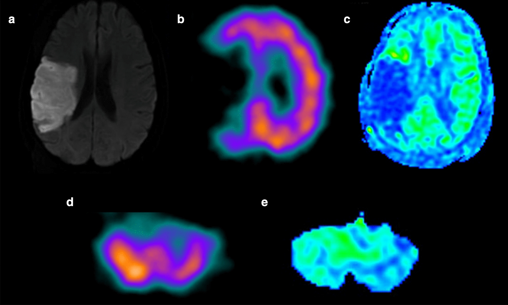
As a noninvasive perfusion-weighted MRI technique, arterial spin-labeling (ASL) was becoming increasingly used to evaluate cerebral hemodynamics in many studies. The relation between ASL-MRI and crossed cerebellar diaschisis (CCD) was rarely discussed. In this study, the aim of our study was to assess the performance of ASL-MRI in the detection of crossed cerebellar diaschisis after stroke in compared with single-photon emission CT (SPECT).

RESULTS

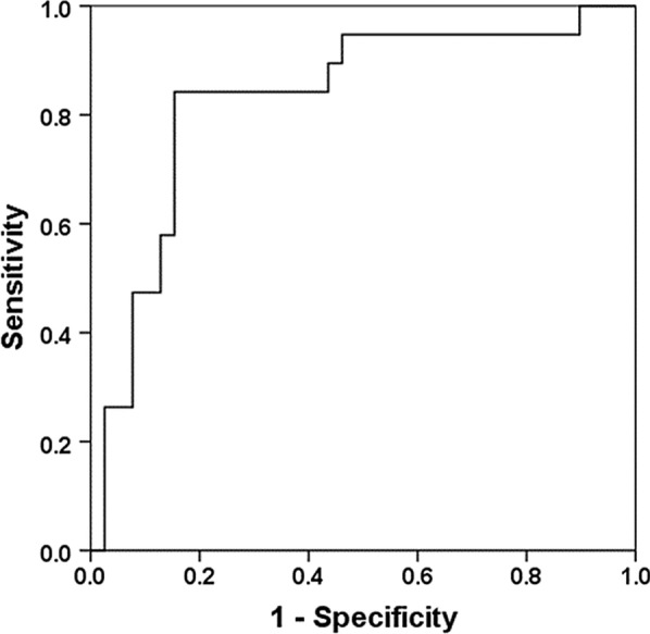
17 of 51(33.3%) patients revealed CCD phenomenon by the SPECT method. In CCD-positive group, CBF of ipsilateral cerebellar were significantly increased compared with contralateral cerebellar (p < 0.0001) while no significant differences (p = 0.063, > 0.001) in the CCD-negative group. Positive correlation was detected between admission National institute of health stroke scale (NIHSS) and asymmetry index of SPECT (AI) (r = 0.351, p = 0.011), AI (r = 0.372, p = 0.007); infract volume and AI (r = 0.443, p = 0.001), AI (r = 0.426, p = 0.002). Significant correlation was also found between cerebral blood flow of SPECT (CBF) and CBF, AI and AI (r = 0.204, p = 0.04; r = 0.467, p = 0.001, respectively). Furthermore, the area under the receiver operating characteristic (ROC) curve value of AI was 0.829.

CONCLUSIONS

CBF derived from ASL-MRI could be valuable for assessment of CCD in supratentorial stroke patients. Additionally, CCD was significantly associated with larger ischemic volume and higher initial NIHSS score.

摘要

背景

动脉自旋标记(ASL)作为一种非侵入性灌注加权 MRI 技术,在许多研究中越来越多地用于评估脑血流动力学。ASL-MRI 与交叉性小脑失联络(CCD)之间的关系很少被讨论。本研究旨在评估 ASL-MRI 在检测脑卒中后交叉性小脑失联络方面的性能,并与单光子发射 CT(SPECT)进行比较。

结果

SPECT 方法发现 51 例患者中有 17 例(33.3%)存在 CCD 现象。在 CCD 阳性组,对侧小脑的 CBF 明显高于同侧小脑(p<0.0001),而在 CCD 阴性组无明显差异(p=0.063,>0.001)。入院时国立卫生研究院卒中量表(NIHSS)评分与 SPECT 不对称指数(AI)之间存在正相关(r=0.351,p=0.011),AI(r=0.372,p=0.007);梗死体积与 AI(r=0.443,p=0.001)、AI(r=0.426,p=0.002)之间存在正相关。SPECT 的 CBF 与 CBF 之间也存在显著相关性、AI 与 AI(r=0.204,p=0.04;r=0.467,p=0.001)。此外,AI 的 ROC 曲线下面积值为 0.829。

结论

ASL-MRI 衍生的 CBF 可用于评估幕上脑卒中患者的 CCD。此外,CCD 与较大的缺血体积和较高的初始 NIHSS 评分显著相关。

https://cdn.ncbi.nlm.nih.gov/pmc/blobs/0d60/7678313/c46c44028635/12868_2020_595_Fig3_HTML.jpg
https://cdn.ncbi.nlm.nih.gov/pmc/blobs/0d60/7678313/9e3af4447545/12868_2020_595_Fig1_HTML.jpg
https://cdn.ncbi.nlm.nih.gov/pmc/blobs/0d60/7678313/a70787fbaa29/12868_2020_595_Fig2_HTML.jpg
https://cdn.ncbi.nlm.nih.gov/pmc/blobs/0d60/7678313/c46c44028635/12868_2020_595_Fig3_HTML.jpg
https://cdn.ncbi.nlm.nih.gov/pmc/blobs/0d60/7678313/9e3af4447545/12868_2020_595_Fig1_HTML.jpg
https://cdn.ncbi.nlm.nih.gov/pmc/blobs/0d60/7678313/a70787fbaa29/12868_2020_595_Fig2_HTML.jpg
https://cdn.ncbi.nlm.nih.gov/pmc/blobs/0d60/7678313/c46c44028635/12868_2020_595_Fig3_HTML.jpg

相似文献

1
Crossed cerebellar diaschisis after stroke detected noninvasively by arterial spin-labeling MR imaging.通过动脉自旋标记磁共振成像无创性检测脑卒中后交叉性小脑 diaschisis。
BMC Neurosci. 2020 Nov 20;21(1):46. doi: 10.1186/s12868-020-00595-z.
2
Detection of Crossed Cerebellar Diaschisis by Intravoxel Incoherent Motion MR Imaging in Subacute Ischemic Stroke.脑桥交叉性小脑失联络的磁共振体素内不相干运动弥散加权成像研究。
Cell Transplant. 2019 Aug;28(8):1062-1070. doi: 10.1177/0963689719856290. Epub 2019 Jun 14.
3
Crossed cerebellar diaschisis detected by arterial spin-labeled perfusion magnetic resonance imaging in subacute ischemic stroke.动脉自旋标记灌注磁共振成像检测亚急性缺血性卒中的交叉性小脑失联络
J Stroke Cerebrovasc Dis. 2014 Oct;23(9):2378-83. doi: 10.1016/j.jstrokecerebrovasdis.2014.05.009. Epub 2014 Aug 31.
4
Crossed cerebellar diaschisis after stroke identified noninvasively with cerebral blood flow-weighted arterial spin labeling MRI.采用脑血流加权动脉自旋标记磁共振成像无创识别卒中后交叉性小脑失联络
Eur J Radiol. 2016 Jan;85(1):136-142. doi: 10.1016/j.ejrad.2015.11.003. Epub 2015 Nov 10.
5
Correlation of Asymmetry Indices Measured by Arterial Spin-Labeling MR Imaging and SPECT in Patients with Crossed Cerebellar Diaschisis.交叉性小脑失联络患者动脉自旋标记磁共振成像和单光子发射计算机断层扫描测量的不对称指数的相关性
AJNR Am J Neuroradiol. 2015 Sep;36(9):1662-8. doi: 10.3174/ajnr.A4366. Epub 2015 Jul 30.
6
Detection of crossed cerebellar diaschisis in hyperacute ischemic stroke using arterial spin-labeled MR imaging.使用动脉自旋标记磁共振成像检测超急性缺血性卒中的交叉性小脑失联络
PLoS One. 2017 Mar 21;12(3):e0173971. doi: 10.1371/journal.pone.0173971. eCollection 2017.
7
Crossed cerebellar diaschisis after acute ischemic stroke detected by intravoxel incoherent motion magnetic resonance imaging.脑桥交叉性小脑失联络现象在急性缺血性卒中中的扩散张量磁共振研究
Neurol Sci. 2022 Feb;43(2):1135-1141. doi: 10.1007/s10072-021-05425-6. Epub 2021 Jul 2.
8
Crossed cerebellar diaschisis-related supratentorial hemodynamic and metabolic status measured by PET/MR in assessing postoperative prognosis in chronic ischemic cerebrovascular disease patients with bypass surgery.采用 PET/MR 测量交叉性小脑 diaschisis 相关的幕上血液动力学和代谢状态评估旁路手术后慢性缺血性脑血管病患者的术后预后。
Ann Nucl Med. 2022 Sep;36(9):812-822. doi: 10.1007/s12149-022-01766-0. Epub 2022 Jul 5.
9
Crossed cerebellar diaschisis in acute stroke detected by dynamic susceptibility contrast MR perfusion imaging.动态磁敏感对比增强磁共振灌注成像检测急性卒中中的交叉性小脑失联络
AJNR Am J Neuroradiol. 2009 Apr;30(4):710-5. doi: 10.3174/ajnr.A1435. Epub 2009 Feb 4.
10
Early-phase 18F-AV-45 PET Imaging can Detect Crossed Cerebellar Diaschisis Following Carotid Artery Stenosis and Cerebral Hypoperfusion.早期18F-AV-45正电子发射断层显像(PET)成像可检测颈动脉狭窄和脑灌注不足后的交叉性小脑失联络。
Curr Neurovasc Res. 2017;14(3):258-265. doi: 10.2174/1567202614666170621102101.

引用本文的文献

1
The power spectrum and functional connectivity characteristics of resting-state EEG in patients with generalized anxiety disorder.广泛性焦虑症患者静息态脑电图的功率谱和功能连接特征
Sci Rep. 2025 Feb 18;15(1):5991. doi: 10.1038/s41598-025-90362-z.
2
Out of the core: the impact of focal ischemia in regions beyond the penumbra.核心之外:半暗带以外区域局灶性缺血的影响
Front Cell Neurosci. 2024 Mar 5;18:1336886. doi: 10.3389/fncel.2024.1336886. eCollection 2024.
3
Disability and persistent motor deficits are linked to structural crossed cerebellar diaschisis in chronic stroke.

本文引用的文献

1
Detection of Crossed Cerebellar Diaschisis by Intravoxel Incoherent Motion MR Imaging in Subacute Ischemic Stroke.脑桥交叉性小脑失联络的磁共振体素内不相干运动弥散加权成像研究。
Cell Transplant. 2019 Aug;28(8):1062-1070. doi: 10.1177/0963689719856290. Epub 2019 Jun 14.
2
Perfusion MR Imaging Using a 3D Pulsed Continuous Arterial Spin-Labeling Method for Acute Cerebral Infarction Classified as Branch Atheromatous Disease Involving the Lenticulostriate Artery Territory.使用三维脉冲连续动脉自旋标记法对累及豆纹动脉区域的分支动脉粥样硬化性疾病所致急性脑梗死进行灌注磁共振成像。
AJNR Am J Neuroradiol. 2017 Aug;38(8):1550-1554. doi: 10.3174/ajnr.A5247. Epub 2017 Jun 8.
3
残疾和持续的运动缺陷与慢性中风中的结构性交叉小脑失联络有关。
Hum Brain Mapp. 2023 Nov;44(16):5336-5345. doi: 10.1002/hbm.26434. Epub 2023 Jul 20.
4
Hyperperfusion in the cerebellum lobule VIIb in patients with epileptic seizures.小脑 VIIb 叶的灌注过度与癫痫发作有关。
BMC Neurol. 2022 Sep 16;22(1):352. doi: 10.1186/s12883-022-02882-0.
5
Progressive Crossed Cerebellar Wallerian Degeneration After Hemispheric Infarct.半球梗死后脑桥交叉性进行性华勒氏变性
Stroke. 2022 Apr;53(4):e143-e144. doi: 10.1161/STROKEAHA.122.038915. Epub 2022 Mar 21.
6
A CBF decrease in the left supplementary motor areas: New insight into postoperative pediatric cerebellar mutism syndrome using arterial spin labeling perfusion MRI.左侧补充运动区 CBF 下降:应用动脉自旋标记灌注 MRI 对术后小儿小脑缄默症的新认识。
J Cereb Blood Flow Metab. 2021 Dec;41(12):3339-3349. doi: 10.1177/0271678X211031321. Epub 2021 Jul 14.
7
Crossed cerebellar diaschisis after acute ischemic stroke detected by intravoxel incoherent motion magnetic resonance imaging.脑桥交叉性小脑失联络现象在急性缺血性卒中中的扩散张量磁共振研究
Neurol Sci. 2022 Feb;43(2):1135-1141. doi: 10.1007/s10072-021-05425-6. Epub 2021 Jul 2.
Detection of crossed cerebellar diaschisis in hyperacute ischemic stroke using arterial spin-labeled MR imaging.
使用动脉自旋标记磁共振成像检测超急性缺血性卒中的交叉性小脑失联络
PLoS One. 2017 Mar 21;12(3):e0173971. doi: 10.1371/journal.pone.0173971. eCollection 2017.
4
Correlation of Asymmetry Indices Measured by Arterial Spin-Labeling MR Imaging and SPECT in Patients with Crossed Cerebellar Diaschisis.交叉性小脑失联络患者动脉自旋标记磁共振成像和单光子发射计算机断层扫描测量的不对称指数的相关性
AJNR Am J Neuroradiol. 2015 Sep;36(9):1662-8. doi: 10.3174/ajnr.A4366. Epub 2015 Jul 30.
5
Two Cases of Crossed Cerebellar Diaschisis with or without Thalamic Lesion on Brain MRI in Status Epilepticus.癫痫持续状态时脑MRI显示有或无丘脑病变的交叉性小脑失联络两例
J Epilepsy Res. 2014 Dec 31;4(2):74-7. doi: 10.14581/jer.14015. eCollection 2014 Dec.
6
Crossed cerebellar diaschisis detected by arterial spin-labeled perfusion magnetic resonance imaging in subacute ischemic stroke.动脉自旋标记灌注磁共振成像检测亚急性缺血性卒中的交叉性小脑失联络
J Stroke Cerebrovasc Dis. 2014 Oct;23(9):2378-83. doi: 10.1016/j.jstrokecerebrovasdis.2014.05.009. Epub 2014 Aug 31.
7
Crossed cerebellar diaschisis due to cerebral diffuse large B cell lymphoma on 18F-FDG PET/CT.18F-FDG PET/CT显示的大脑弥漫性大B细胞淋巴瘤所致的交叉性小脑失联络
Int J Hematol. 2014 Nov;100(5):415-6. doi: 10.1007/s12185-014-1656-1. Epub 2014 Aug 14.
8
Concurrence of crossed cerebellar diaschisis and parakinesia brachialis oscitans in a patient with hemorrhagic stroke.一名出血性中风患者出现交叉性小脑性共济失调和哈欠样臂部运动障碍并存的情况。
Case Rep Med. 2013;2013:519808. doi: 10.1155/2013/519808. Epub 2013 Nov 6.
9
Recurrent glioma and crossed cerebellar diaschisis in a patient examined with 18F-DOPA and 18F-FDG PET/CT.患者经 18F-DOPA 和 18F-FDG PET/CT 检查后发现复发性脑胶质瘤和交叉性小脑失联络。
Clin Nucl Med. 2012 Sep;37(9):878-9. doi: 10.1097/RLU.0b013e318262af2a.
10
Crossed cerebro-cerebellar diaschisis.交叉性大脑小脑失联络
Neuropediatrics. 2012 Apr;43(2):53-4. doi: 10.1055/s-0032-1310543. Epub 2012 Apr 3.