Tang Liqun, Xie Jianhong, Yu Xiaoqin, Zheng Yangyang
Department of Geriatrics, Zhejiang Province People's Hospital, Hangzhou Medical College, Hangzhou, Zhejiang, China.
Department of Geriatrics, Zhejiang Aid Hospital, Hangzhou, Zhejiang, China.
PeerJ. 2020 Nov 17;8:e10371. doi: 10.7717/peerj.10371. eCollection 2020.
The role of miR-26a-5p expression in cardiac hypertrophy remains unclear. Herein, the effect of miR-26a-5p on cardiac hypertrophy was investigated using phenylephrine (PE)-induced cardiac hypertrophy in vitro and in a rat model of hypertension-induced hypertrophy in vivo.
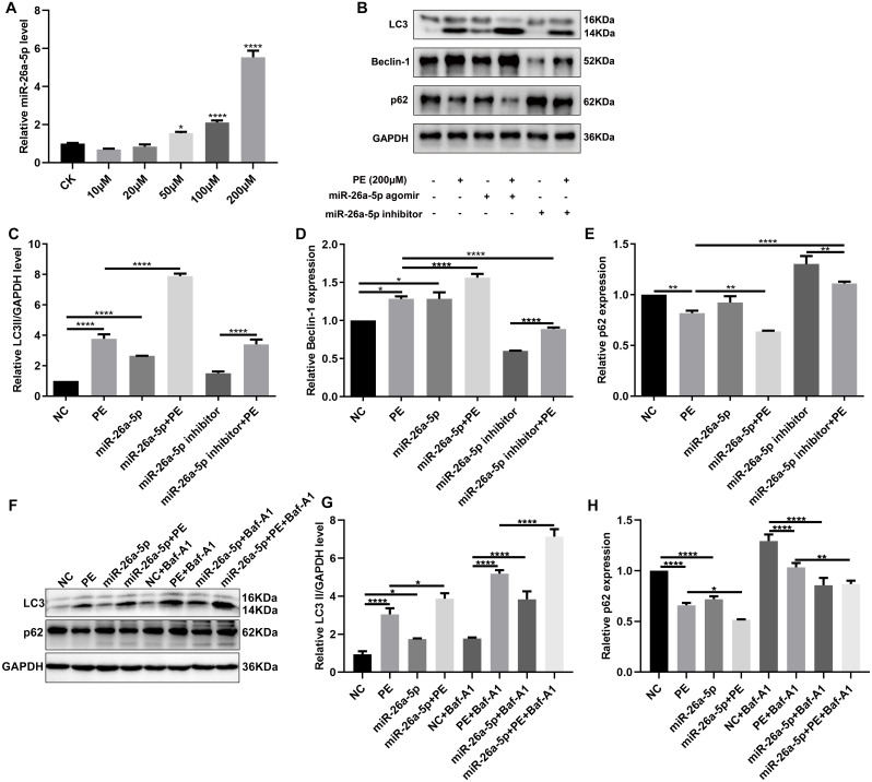
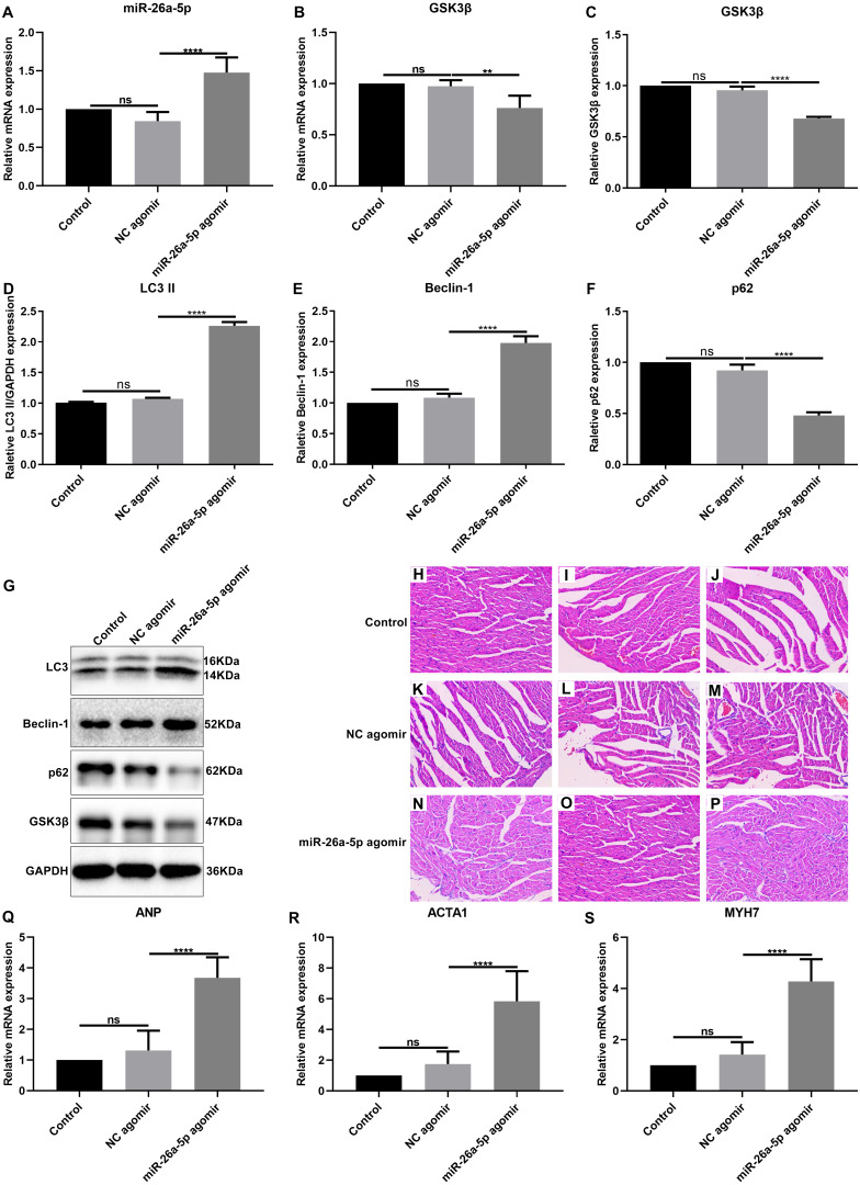
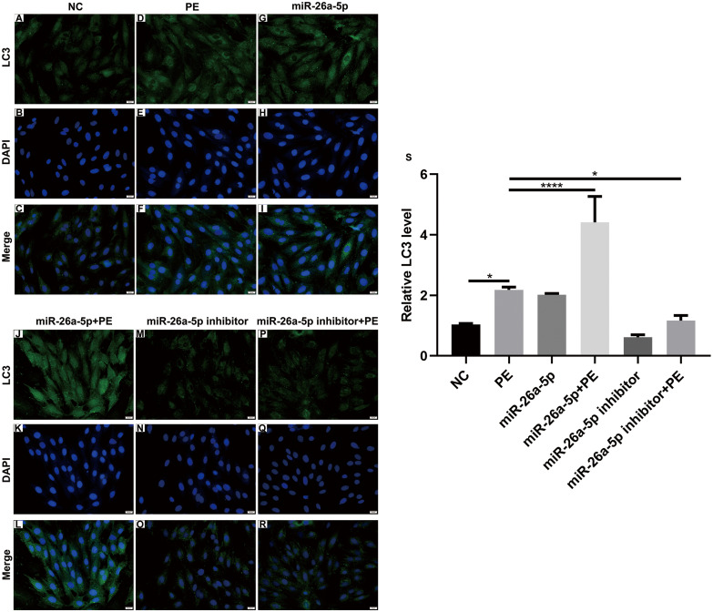
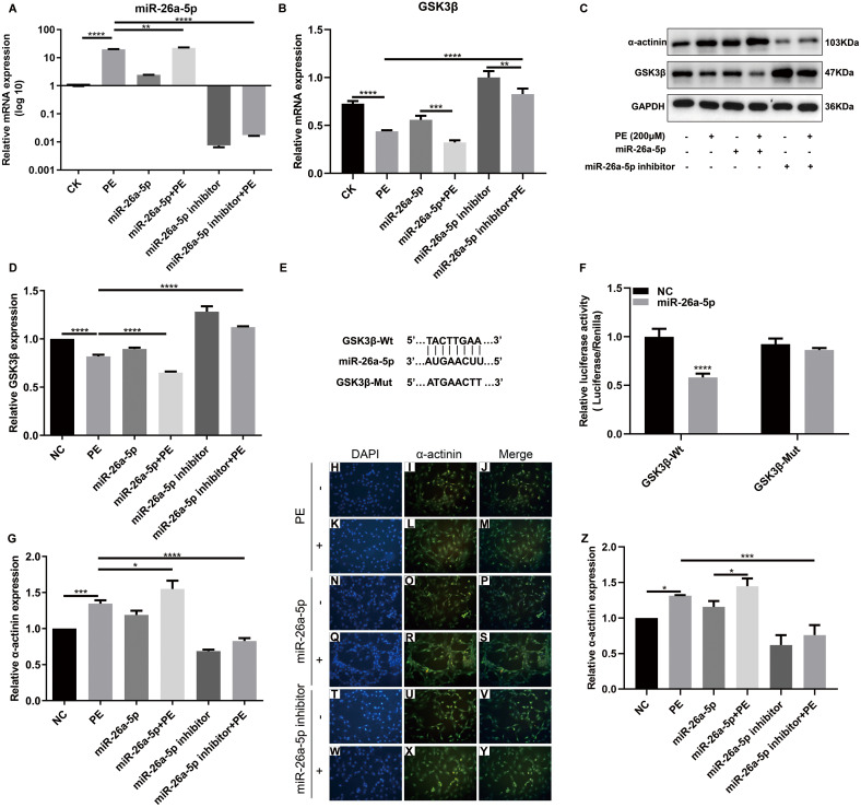
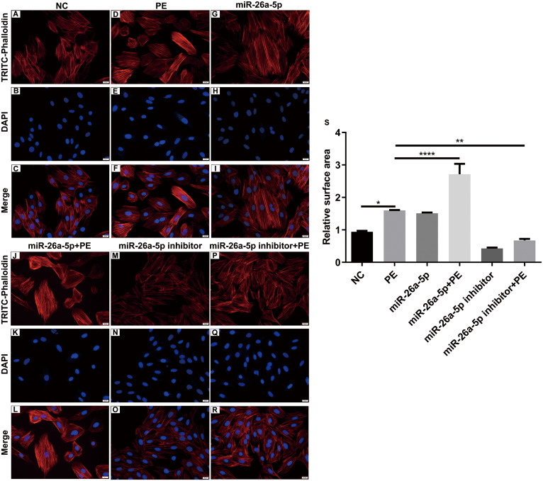
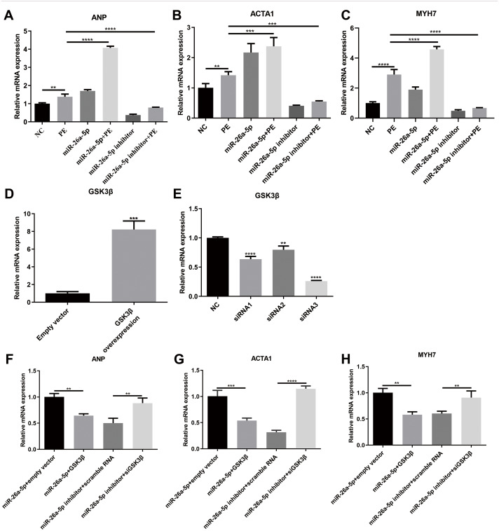
The PE-induced cardiac hypertrophy models in vitro and vivo were established. To investigate the effect of miR-26a-5p activation on autophagy, the protein expression of autophagosome marker (LC3) and p62 was detected by western blot analysis. To explore the effect of miR-26a-5p activation on cardiac hypertrophy, the relative mRNA expression of cardiac hypertrophy related mark GSK3 was detected by qRT-PCR in vitro and vivo. In addition, immunofluorescence staining was used to detect cardiac hypertrophy related mark -actinin. The cell surface area was measured by immunofluorescence staining. The direct target relationship between miR-26a-5p and GSK3 was confirmed by dual luciferase report.
MiR-26a-5p was highly expressed in PE-induced cardiac hypertrophy. MiR-26a-5p promoted LC3II and decreased p62 expression in PE-induced cardiac hypertrophy in the presence or absence of lysosomal inhibitor. Furthermore, miR-26a-5p significantly inhibited GSK3 expression in vitro and in vivo. Dual luciferase report results confirmed that miR-26a-5p could directly target GSK3. GSK3 overexpression significantly reversed the expression of cardiac hypertrophy-related markers including ANP, ACTA1 and MYH7. Immunofluorescence staining results demonstrated that miR-26a-5p promoted cardiac hypertrophy related protein -actinin expression, and increased cell surface area in vitro and in vivo.
Our study revealed that miR-26a-5p promotes myocardial cell autophagy activation and cardiac hypertrophy by regulating GSK3, which needs further research.
miR-26a-5p表达在心肌肥厚中的作用仍不清楚。在此,利用苯肾上腺素(PE)诱导的体外心肌肥厚以及体内高血压诱导的肥厚大鼠模型,研究了miR-26a-5p对心肌肥厚的影响。
建立了PE诱导的体外和体内心肌肥厚模型。为研究miR-26a-5p激活对自噬的影响,通过蛋白质免疫印迹分析检测自噬体标志物(LC3)和p62的蛋白表达。为探究miR-26a-5p激活对心肌肥厚的影响,通过实时定量聚合酶链反应(qRT-PCR)检测体外和体内心肌肥厚相关标志物GSK3的相对mRNA表达。此外,采用免疫荧光染色检测心肌肥厚相关标志物α-辅肌动蛋白。通过免疫荧光染色测量细胞表面积。通过双荧光素酶报告实验证实miR-26a-5p与GSK3之间的直接靶向关系。
miR-26a-5p在PE诱导的心肌肥厚中高表达。在存在或不存在溶酶体抑制剂的情况下,miR-26a-5p均促进PE诱导的心肌肥厚中LC3II表达并降低p62表达。此外,miR-26a-5p在体外和体内均显著抑制GSK3表达。双荧光素酶报告实验结果证实miR-26a-5p可直接靶向GSK3。GSK3过表达显著逆转了包括心钠素(ANP)、α-平滑肌肌动蛋白(ACTA1)和肌球蛋白重链7(MYH7)在内的心肌肥厚相关标志物的表达。免疫荧光染色结果表明,miR-26a-5p促进体外和体内心肌肥厚相关蛋白α-辅肌动蛋白表达,并增加细胞表面积。
我们的研究表明,miR-26a-5p通过调节GSK3促进心肌细胞自噬激活和心肌肥厚,这有待进一步研究。