Department of Biomedical Science, CHA University, Seongnam, 13488, Republic of Korea.
Department of Oral Pathology, College of Dentistry, Gangneung-Wonju National University, Gangneung, 25457, Republic of Korea.
Stem Cell Res Ther. 2020 Nov 27;11(1):512. doi: 10.1186/s13287-020-02029-3.
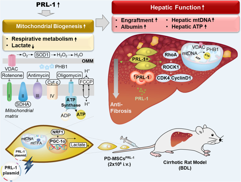
Placenta-derived mesenchymal stem cells (PD-MSCs) have been highlighted as an alternative cell therapy agent that has become a next-generation stem cell treatment. Phosphatase of regenerating liver-1 (PRL-1), an immediate early gene, plays a critical role during liver regeneration. Here, we generated enhanced PRL-1 in PD-MSCs (PD-MSCs, PRL-1+) using lentiviral and nonviral gene delivery systems and investigated mitochondrial functions by PD-MSC transplantation for hepatic functions in a rat bile duct ligation (BDL) model.
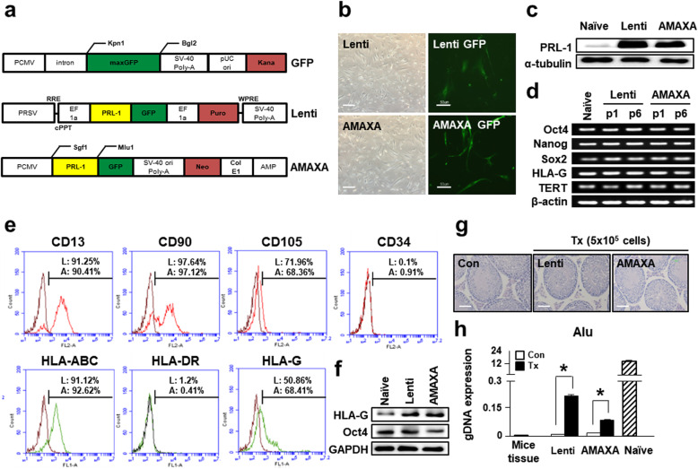
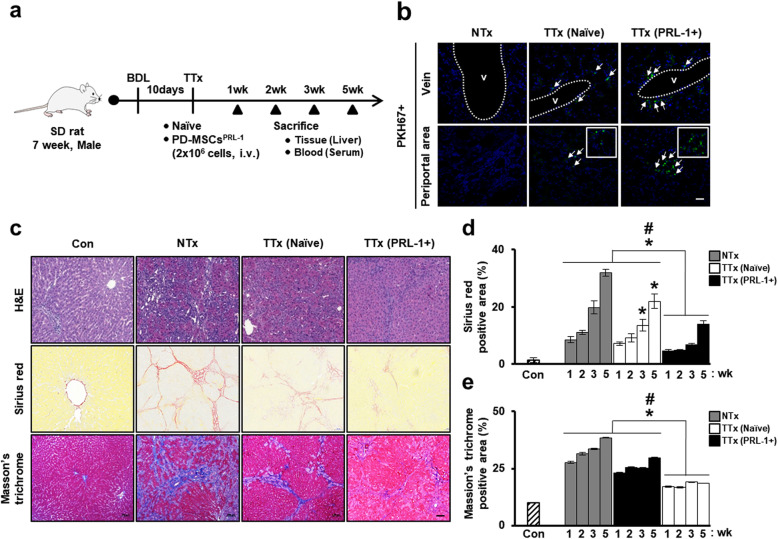
PD-MSCs were generated by lentiviral and nonviral AMAXA gene delivery systems and analyzed for their characteristics and mitochondrial metabolic functions. Liver cirrhosis was induced in Sprague-Dawley (SD) rats using common BDL for 10 days. PKH67+ naïve and PD-MSCs using a nonviral sysyem (2 × 10 cells/animal) were intravenously administered into cirrhotic rats. The animals were sacrificed at 1, 2, 3, and 5 weeks after transplantation and engraftment of stem cells, and histopathological analysis and hepatic mitochondrial functions were performed.
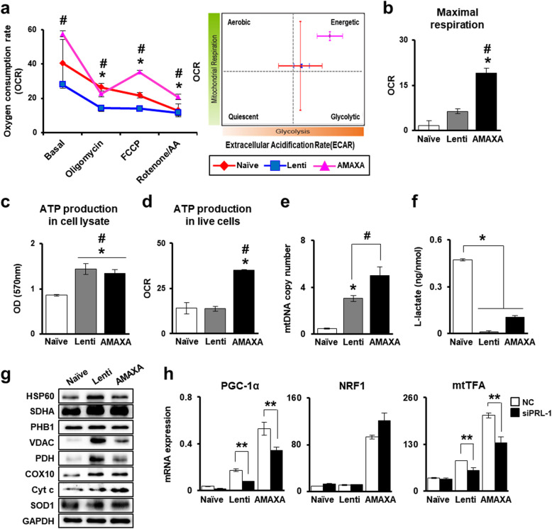
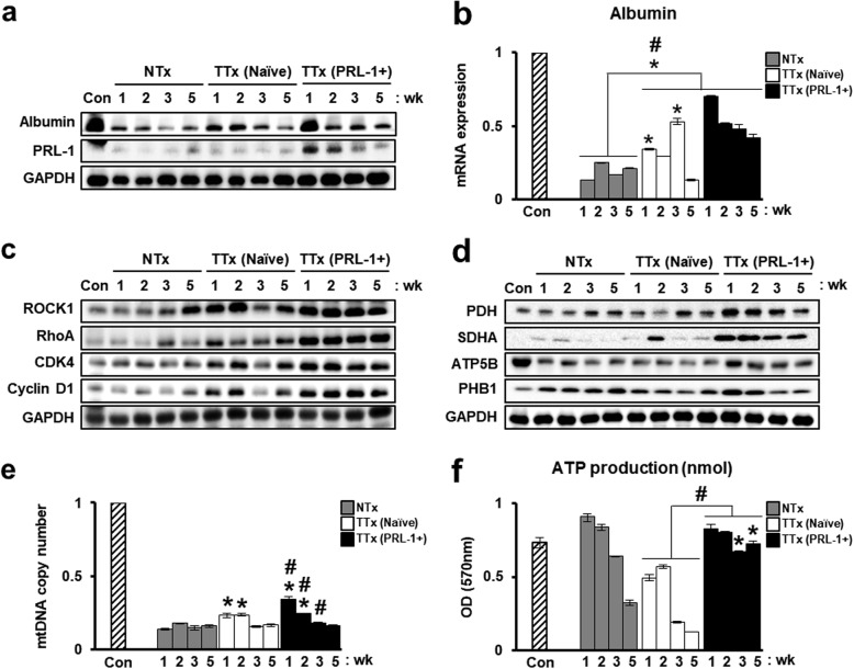
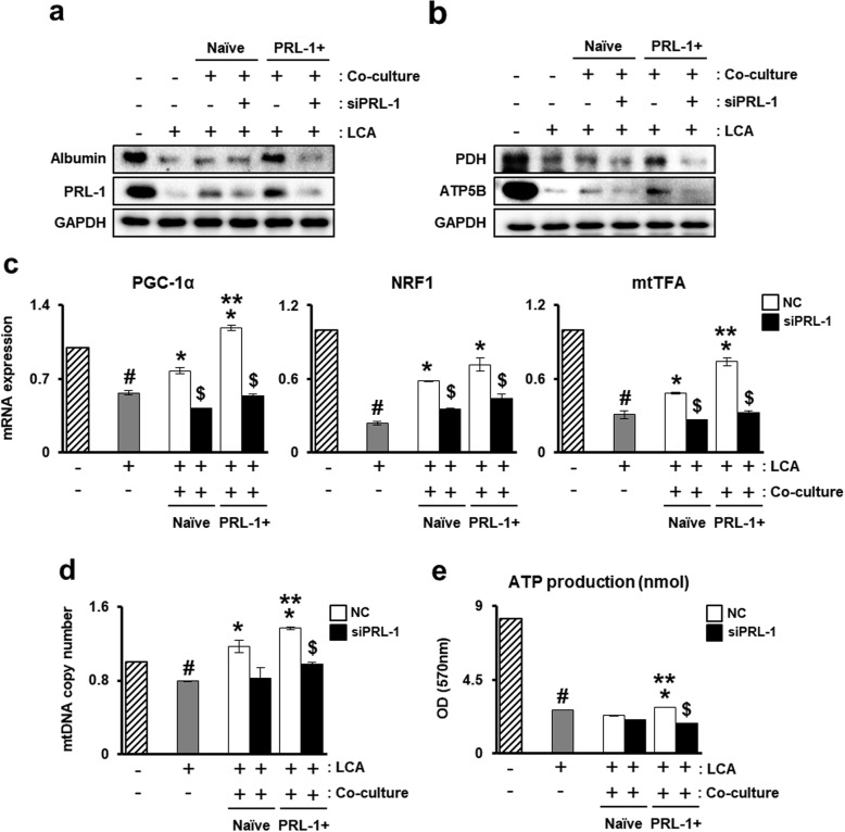
PD-MSCs were successfully generated using lentiviral and nonviral AMAXA systems and maintained characteristics similar to those of naïve cells. Compared with naïve cells, PD-MSCs improved respirational metabolic states of mitochondria. In particular, mitochondria in PD-MSCs generated by the nonviral AMAXA system showed a significant increase in the respirational metabolic state, including ATP production and mitochondrial biogenesis (*p < 0.05). Furthermore, transplantation of PD-MSCs using a nonviral AMAXA system promoted engraftment into injured target liver tissues of a rat BDL cirrhotic model and enhanced the metabolism of mitochondria via increased mtDNA and ATP production, thereby improving therapeutic efficacy.
Our findings will further our understanding of the therapeutic mechanism of enhanced MSCs and provide useful data for the development of next-generation MSC-based cell therapy and therapeutic strategies for regenerative medicine in liver disease.
胎盘间充质干细胞(PD-MSCs)已被强调为一种替代细胞治疗剂,成为下一代干细胞治疗方法。肝再生磷酸酶-1(PRL-1)是一种即刻早期基因,在肝再生过程中发挥关键作用。在这里,我们使用慢病毒和非病毒基因传递系统在 PD-MSCs 中产生增强的 PRL-1(PD-MSCs,PRL-1+),并通过 PD-MSC 移植在大鼠胆管结扎(BDL)模型中研究线粒体功能对肝功能的影响。
使用慢病毒和非病毒 AMAXA 基因传递系统生成 PD-MSCs,并分析其特征和线粒体代谢功能。使用普通 BDL 在 Sprague-Dawley(SD)大鼠中诱导肝硬化 10 天。将 PKH67+幼稚和使用非病毒系统的 PD-MSCs(每只动物 2×10 个细胞)静脉内给予肝硬化大鼠。在移植后 1、2、3 和 5 周时处死动物,并进行组织病理学分析和肝线粒体功能检测。
成功使用慢病毒和非病毒 AMAXA 系统生成 PD-MSCs,并保持与幼稚细胞相似的特征。与幼稚细胞相比,PD-MSCs 改善了线粒体的呼吸代谢状态。特别是,非病毒 AMAXA 系统产生的 PD-MSCs 的线粒体呼吸代谢状态显著增加,包括 ATP 产生和线粒体生物发生(*p<0.05)。此外,非病毒 AMAXA 系统移植 PD-MSCs 促进了大鼠 BDL 肝硬化模型受损靶肝组织的植入,并通过增加 mtDNA 和 ATP 产生来增强线粒体代谢,从而提高治疗效果。
我们的研究结果将进一步加深我们对增强型 MSCs 治疗机制的理解,并为下一代基于 MSC 的细胞治疗和肝脏疾病再生医学的治疗策略的发展提供有用的数据。