van Oirschot Bart A J A, Jansen John A, van de Ven Cindy J J M, Geven Edwin J W, Gossen Jan A
Department of Dentistry - Biomaterials, Radboudumc, Radboud University Nijmegen, NijmegenThe Netherlands.
Osteo-Pharma BV, OssThe Netherlands.
J Oral Maxillofac Res. 2020 Nov 30;11(3):e4. doi: 10.5037/jomr.2020.11304. eCollection 2020 Jul-Sep.
The purpose of the present study was to evaluate whether pericard collagen membranes coated with ancillary amounts of testosterone and alendronate in a poly-lactic glycolic acid (PLGA) carrier as compared to uncoated membranes will improve early bone regeneration.
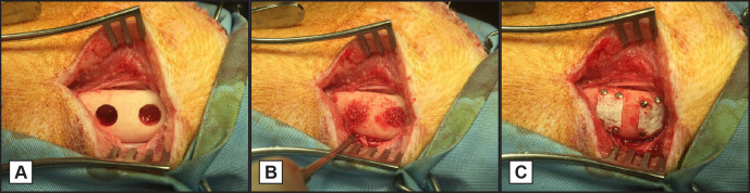
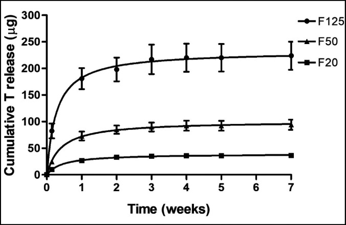
In each of 16 minipigs, four standardized mandibular intraosseous defects were made bilaterally. The defects were filled with Bio-Oss granules and covered with a non-coated or coated membrane. Membranes were spray-coated with 4 layers of PLGA containing testosterone and alendronate resulting in 20, 50 or 125 μg/cm of testosterone and 20 µg/cm alendronate (F20, F50, F125). Non-coated membranes served as controls (F0). Animals were sacrificed at 6 and 12 weeks after treatment. Qualitative and quantitative histological evaluations of bone regeneration were performed. Differences between groups were assessed by paired Student's t-test.
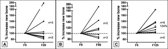
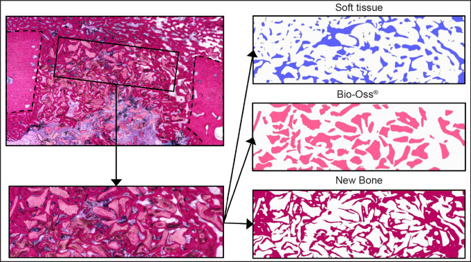
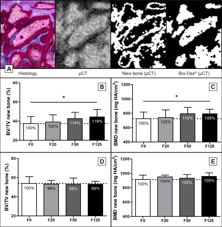
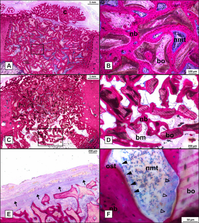
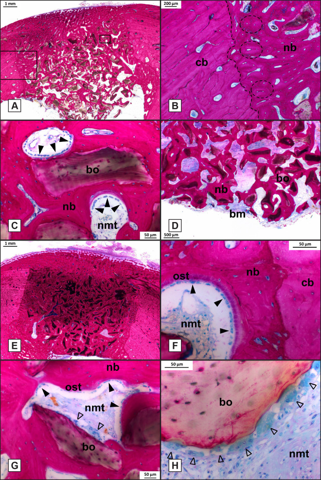
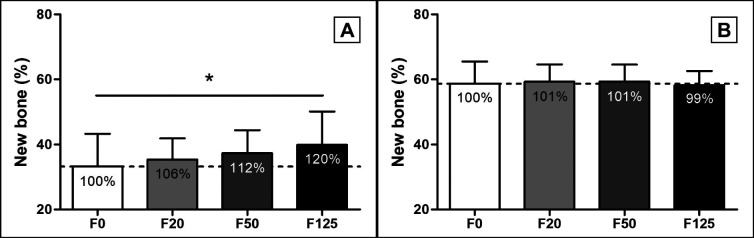
Light microscopical analysis showed new bone formation that was in close contact with the Bio-Oss surface without an intervening non-mineralized tissue layer. Histomorphometric analysis of newly formed bone showed a significant 20% increase in area in the F125 coated membrane treated defects (40 [SD 10]%) compared to the F0 treated defects after 6 weeks (33 [SD 10]%, P = 0.013). At week 12, the total percentage of new bone was increased compared to week 6, but no increase in newly formed bone compared to F0 was observed.
The data from this study indicate that F125 collagen membranes coated with testosterone and alendronate resulted in superior bone formation (+24%) when normalized to control sites using uncoated membranes.
本研究旨在评估与未涂层的心包胶原膜相比,在聚乳酸乙醇酸(PLGA)载体中涂覆辅助量睾酮和阿仑膦酸盐的心包胶原膜是否能改善早期骨再生。
在16只小型猪中,每只双侧制作4个标准化的下颌骨骨内缺损。缺损处填充Bio-Oss颗粒,并用未涂层或涂层的膜覆盖。膜用含睾酮和阿仑膦酸盐的4层PLGA进行喷涂,使睾酮含量达到20、50或125μg/cm,阿仑膦酸盐含量为20μg/cm(F20、F50、F125)。未涂层的膜作为对照(F0)。在治疗后6周和12周处死动物。对骨再生进行定性和定量组织学评估。组间差异采用配对t检验进行评估。
光学显微镜分析显示新骨形成与Bio-Oss表面紧密接触,无中间非矿化组织层。新形成骨的组织形态计量学分析显示,与F0处理的缺损相比,F125涂层膜处理的缺损在6周后新骨面积显著增加20%(40[标准差10]%对33[标准差10]%,P = 0.013)。在第12周,与第6周相比,新骨的总百分比增加,但与F0相比,新形成骨没有增加。
本研究数据表明,与使用未涂层膜的对照部位相比,涂覆睾酮和阿仑膦酸盐的F125胶原膜导致骨形成更优(增加24%)。