Sasaki Shin, Takami Yuko, Wada Yoshiyuki, Ryu Tomoki, Imamura Hajime, Ureshino Hiroki, Fujiwara Minako, Saitsu Hideki
Department of Hepato-Biliary-Pancreatic Surgery, Clinical Research Institute, National Hospital Organization Kyushu Medical Center, 1-8-1 Jigyohama, Chuo-ku, Fukuoka, 810-8563, Japan.
Department of Pathology, National Hospital Organization Kyushu Medical Center, 1-8-1 Jigyohama, Chuo-ku, Fukuoka, 810-8563, Japan.
Surg Case Rep. 2020 Dec 3;6(1):305. doi: 10.1186/s40792-020-01084-5.
Glomus tumors (GTs) are mesenchymal neoplastic lesions arising from the glomus bodies and generally occur in the fingers and toes. Gastrointestinal GTs are rare, and most of them originate from the stomach; however, GT arising from the duodenum is exceedingly rare.
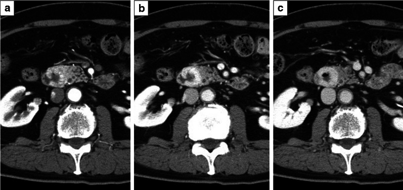
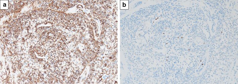
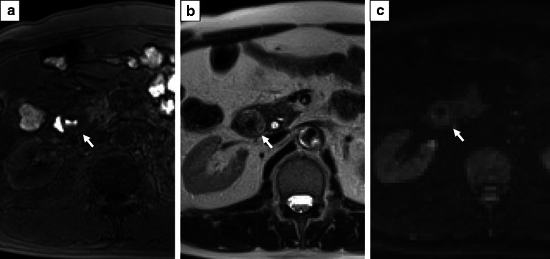
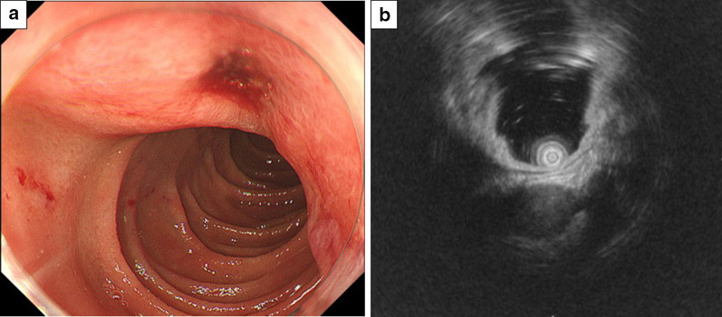
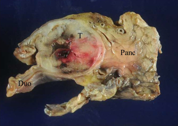
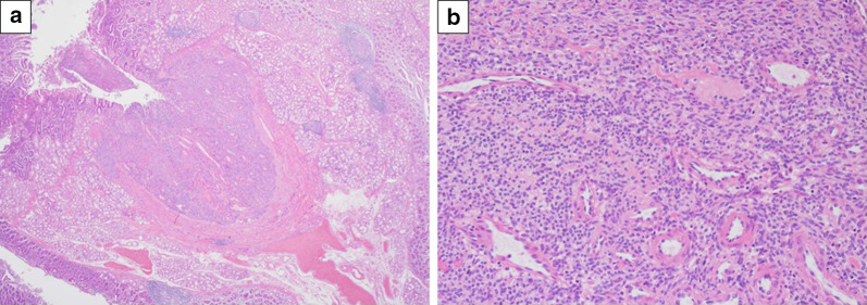
A 68-year-old man was admitted due to abdominal pain. Endoscopy showed a round, smooth, elevated mass in the second portion of the duodenum with central ulceration. Abdominal contrast computed tomography showed a hypervascular tumor measuring 26 mm in diameter in the second portion of the duodenum, and pancreatic invasion was suspected. Endoscopic ultrasonography of the lesion confirmed a hypoechoic mass arising from the fourth layer of the duodenal wall. A biopsy was performed for central ulceration, and immunochemical studies showed positive results for smooth muscle actin (SMA) and negative results for S100, C-Kit, and CD34. Leiomyoma or gastrointestinal stromal tumor was suspected and pancreatoduodenectomy was performed. The specimen exhibited a vascular-rich tumor, 24 × 24 × 19 mm in size, with deep ulceration in the duodenum. Histological examination showed uniform small round cells with central nuclei and a pale cytoplasm (glomus cell) with perivascular proliferation. Immunochemical studies showed that the tumor was positive for SMA and collagen type IV, and negative for C-Kit, CD34, desmin, and S100. We diagnosed the tumor as a GT of the duodenum.
GTs of the duodenum are exceedingly rare, but should be considered in the differential diagnoses of duodenal submucosal lesions.
血管球瘤(GTs)是起源于血管球的间叶性肿瘤性病变,通常发生于手指和脚趾。胃肠道血管球瘤罕见,其中大多数起源于胃;然而,起源于十二指肠的血管球瘤极为罕见。
一名68岁男性因腹痛入院。内镜检查显示十二指肠第二部有一个圆形、光滑、隆起的肿物,中央有溃疡。腹部增强计算机断层扫描显示十二指肠第二部有一个直径26毫米的高血运肿瘤,怀疑侵犯胰腺。对该病变进行内镜超声检查,证实为起源于十二指肠壁第四层的低回声肿物。对中央溃疡进行活检,免疫化学研究显示平滑肌肌动蛋白(SMA)呈阳性,而S100、C-Kit和CD34呈阴性。怀疑为平滑肌瘤或胃肠道间质瘤,遂行胰十二指肠切除术。标本显示为一个富含血管的肿瘤,大小为24×24×19毫米,十二指肠有深度溃疡。组织学检查显示细胞为均匀的小圆形,细胞核位于中央,细胞质淡染(血管球细胞),血管周围有增生。免疫化学研究显示该肿瘤SMA和IV型胶原呈阳性,而C-Kit、CD34、结蛋白和S100呈阴性。我们将该肿瘤诊断为十二指肠血管球瘤。
十二指肠血管球瘤极为罕见,但在十二指肠黏膜下病变的鉴别诊断中应予以考虑。