Zaouak Yasmine, Sadeghi Niloufar, Sarbu Nicolae, Ligot Noémie, Lubicz Boris
Erasme Hospital, BE.
J Belg Soc Radiol. 2020 Nov 25;104(1):70. doi: 10.5334/jbsr.2083.
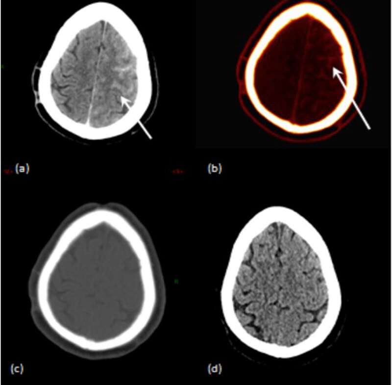
To evaluate the value of dual-energy computed tomography (DECT) in differentiating cerebral hemorrhage from blood brain barrier (BBB) disruption after neuro-interventional procedures with intra-arterial injection of iodinated contrast material.
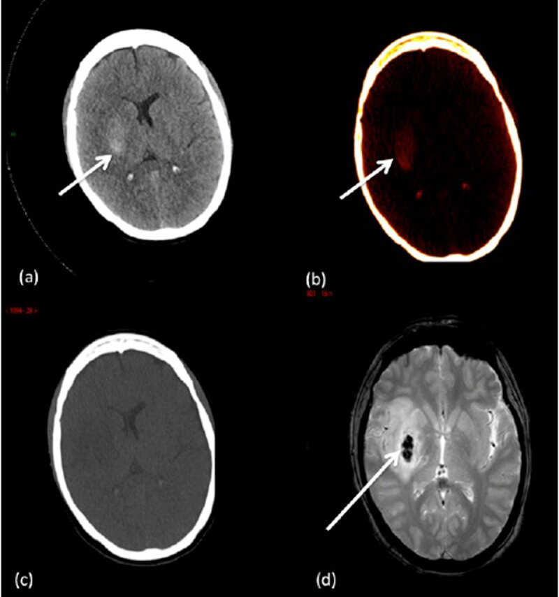
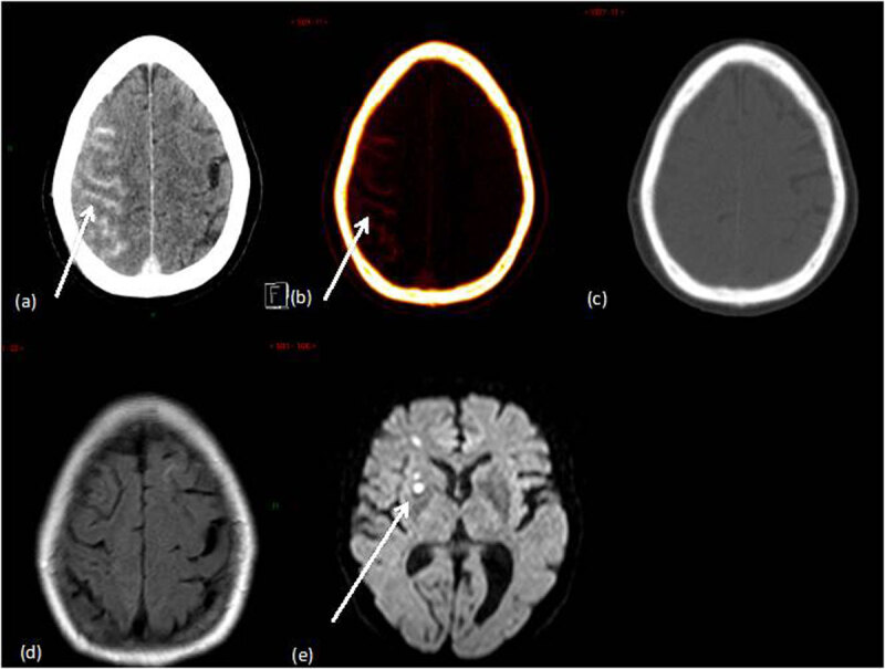
This prospective study was approved by the local ethics committee, and informed consent was obtained for all patients. Thirty five patients with acute ischemic stroke or un-ruptured brain aneurysm who had received intra-arterial administration of iodinated contrast material were evaluated using DECT at 80 and 150 kV immediately after the procedure.A three-material decomposition algorithm was used to obtain virtual non-contrast (VNC) images and iodine overlay maps (IOM). A follow-up examination (brain magnetic resonance imaging MRI or conventional CT) was used as the standard of reference for hemorrhage, defined as a persistant hyperdensity on a conventional CT or T2* hypo-intensity on brain MRI. The diagnostic values of DECT in differentiating hemorrhage and iodinated contrast material were obtained.
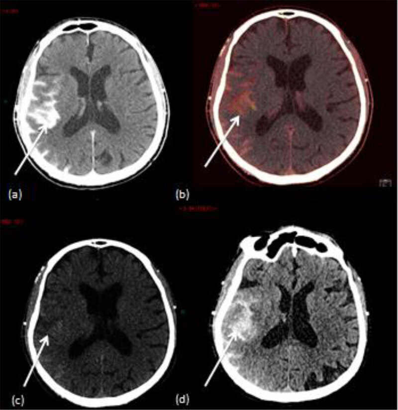
Mixed images obtained with DECT showed intra-parenchymal or subarachnoid hyperattenuation in 18/35 patients. Among these, 16 were classified (according to VNC images and IOM) as contrast extravasations and two with a mixture of hemorrhage and contrast material. On follow-up imaging, there were two patients with hemorrhage. The sensitivity, specificity, and accuracy of DECT in the identifying hemorrhage was calculated as 67% (2/3), 100% (32/32) and 97% (32/33) respectively.
DECT allows an early and accurate differentiation between cerebral hemorrhage and BBB disruption after intra-arterial neuro-interventional procedures.
评估双能计算机断层扫描(DECT)在经动脉注射碘化造影剂的神经介入手术后鉴别脑出血与血脑屏障(BBB)破坏的价值。
本前瞻性研究经当地伦理委员会批准,所有患者均签署知情同意书。对35例接受动脉内注射碘化造影剂的急性缺血性卒中或未破裂脑动脉瘤患者在术后立即采用80 kV和150 kV的DECT进行评估。采用三物质分解算法获得虚拟平扫(VNC)图像和碘叠加图(IOM)。将后续检查(脑磁共振成像MRI或传统CT)作为出血的参考标准,出血定义为传统CT上持续的高密度或脑MRI上的T2*低信号。获得DECT鉴别出血和碘化造影剂的诊断价值。
DECT获得的混合图像显示18/35例患者脑实质内或蛛网膜下腔高密度影。其中,16例(根据VNC图像和IOM)分类为造影剂外渗,2例为出血与造影剂混合。在后续成像中,有2例患者出现出血。DECT识别出血的敏感性、特异性和准确性分别计算为67%(2/3)、100%(32/32)和97%(32/33)。
DECT能够在动脉内神经介入手术后早期准确鉴别脑出血与BBB破坏。