College of Health Solutions, Arizona State University, Phoenix, AZ, USA.
VA Phoenix Health Care System, Phoenix, AZ, USA.
J Int Neuropsychol Soc. 2021 Aug;27(7):733-743. doi: 10.1017/S135561772000123X. Epub 2020 Dec 9.
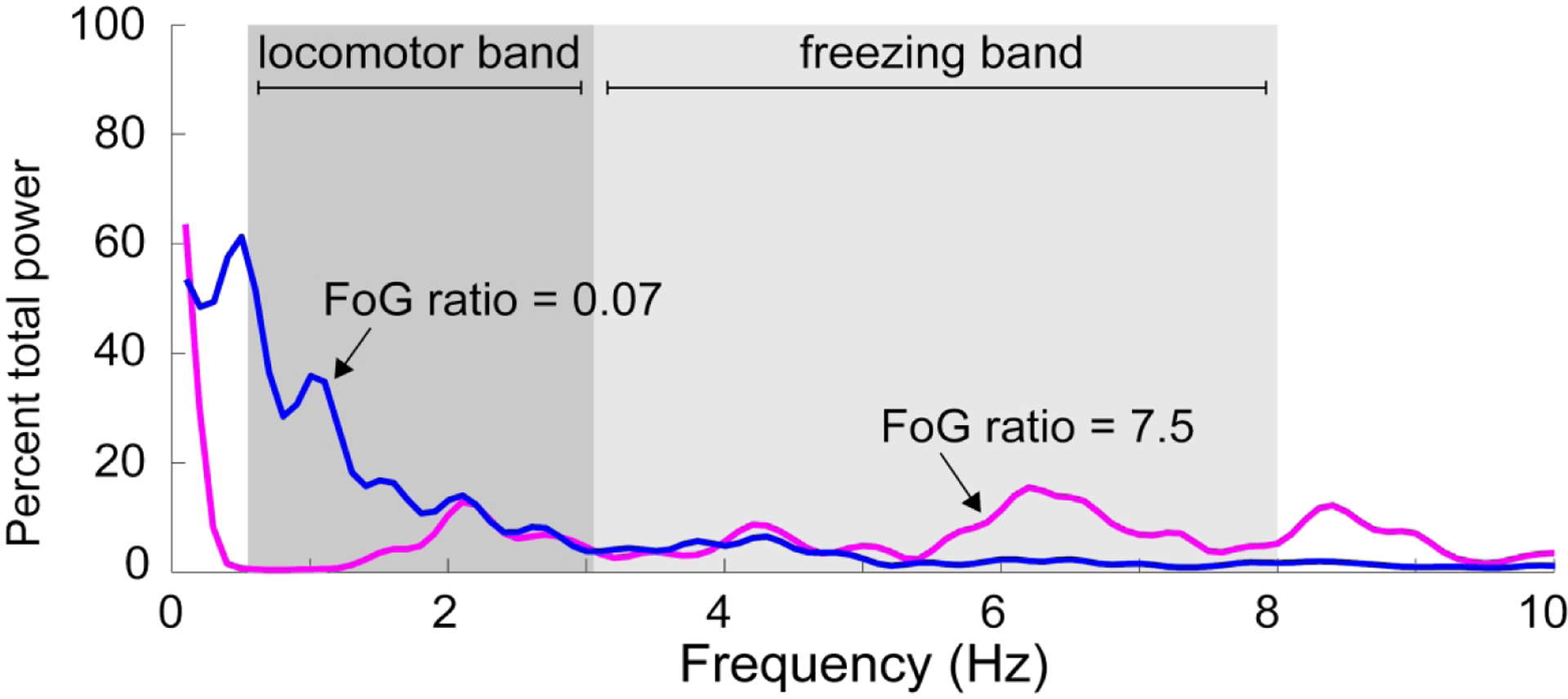
Freezing of gait (FoG) in Parkinson's disease (PD) has been associated with response inhibition. However, the relationship between response inhibition, neural dysfunction, and PD remains unclear. We assessed response inhibition and microstructural integrity of brain regions involved in response inhibition [right hemisphere inferior frontal cortex (IFC), bilateral pre-supplementary motor areas (preSMA), and subthalamic nuclei (STN)] in PD subjects with and without FoG and elderly controls.
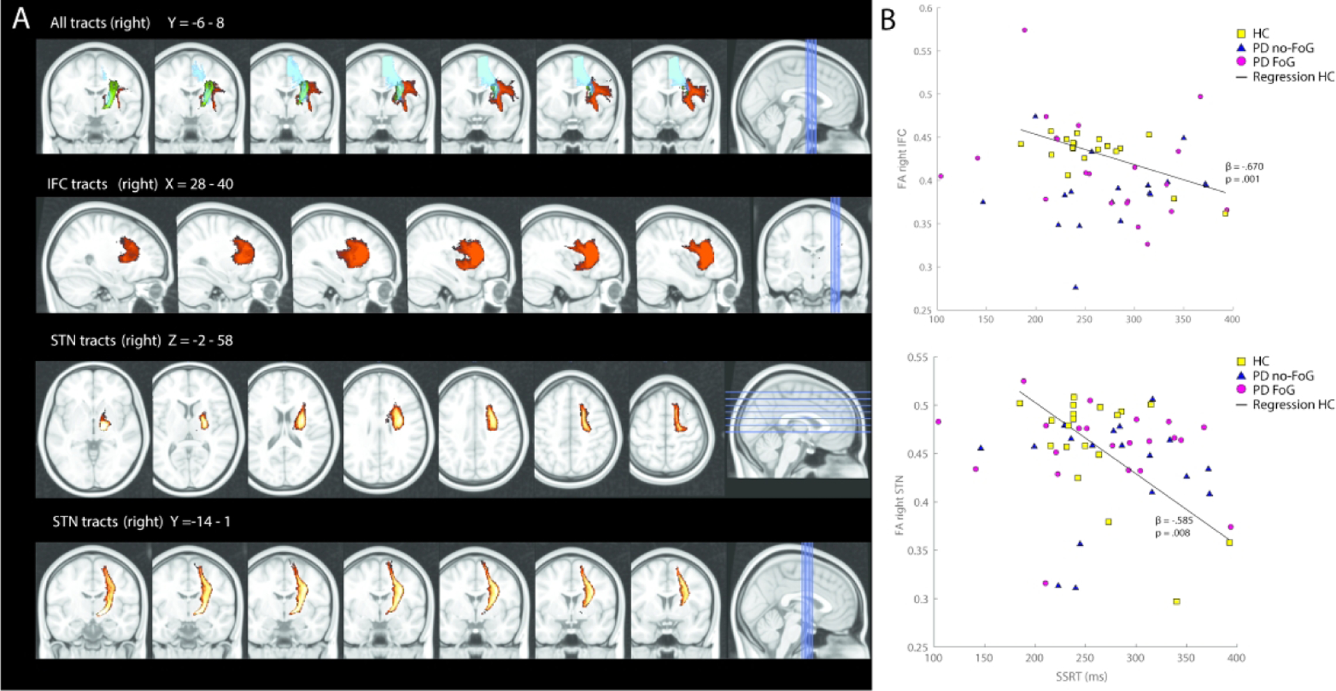
Twenty-one people with PD and FoG (PD-FoG), 18 without FoG (PD-noFoG), and 19 age-matched controls (HC) completed a Stop-Signal Task (SST) and MRI scan. Probabilistic fiber tractography assessed structural integrity (fractional anisotropy, FA) among IFC, preSMA, and STN regions.
Stop-signal performance did not differ between PD and HC, nor between PD-FoG and PD-noFoG. Differences in white matter integrity were observed across groups (.001 < p < .064), but were restricted to PD versus HC groups; no differences in FA were observed between PD-FoG and PD-noFoG (p > .096). Interestingly, worse FoG was associated with higher (better) mean FA in the r-preSMA, (β = .547, p = .015). Microstructural integrity of the r-IFC, r-preSMA, and r-STN tracts correlated with stop-signal performance in HC (p ≤ .019), but not people with PD.
These results do not support inefficient response inhibition in PD-FoG. Those with PD exhibited white matter loss in the response inhibition network, but this was not associated with FoG, nor with response inhibition deficits, suggesting FoG-specific neural changes may occur outside the response inhibition network. As shown previously, white matter loss was associated with response inhibition in elderly controls, suggesting PD may disturb this relationship.
冻结步态(FoG)与帕金森病(PD)的反应抑制有关。然而,反应抑制、神经功能障碍与 PD 之间的关系尚不清楚。我们评估了 PD 伴有和不伴有 FoG 患者以及老年对照组的反应抑制相关脑区[右半球额下回(IFC)、双侧辅助运动前区(preSMA)和丘脑底核(STN)]的反应抑制和微观结构完整性。
21 名 PD 伴 FoG(PD-FoG)患者、18 名无 FoG(PD-noFoG)患者和 19 名年龄匹配的对照组(HC)完成了停止信号任务(SST)和 MRI 扫描。概率纤维束追踪评估了 IFC、preSMA 和 STN 区域的结构完整性(各向异性分数,FA)。
PD 与 HC 之间,以及 PD-FoG 与 PD-noFoG 之间,停止信号的表现均无差异。各组之间存在白质完整性差异(0.001 < p <.064),但仅限于 PD 与 HC 组;PD-FoG 与 PD-noFoG 之间的 FA 无差异(p >.096)。有趣的是,更严重的 FoG 与 r-preSMA 中的更高(更好)平均 FA 相关(β=.547,p =.015)。HC 中 r-IFC、r-preSMA 和 r-STN 束的微观结构完整性与停止信号表现相关(p ≤.019),但 PD 患者中则不相关。
这些结果不支持 PD-FoG 中反应抑制效率低下。PD 患者的反应抑制网络中存在白质丢失,但这与 FoG 无关,也与反应抑制缺陷无关,这表明 FoG 可能发生在反应抑制网络之外的特定神经变化。如前所述,白质丢失与老年对照组的反应抑制有关,这表明 PD 可能干扰了这种关系。