Bravo-Tsri Akoli Eklou Baudouin, Konaté Issa, Kouassi Kouamé Paul Bonfils, Acko-Ohui Estelle Valérie, Goulé-Bi Ange Roland, Isart Dominique, Tanoh Emile Kesse, Vangah Marius Koffi, Kouadio Florent Allou, Yao B L
Radiology Department, Bouaké University Hospital, 01 BP 1174 Bouaké 01, 00225 Bouaké, Ivory Coast.
Radiology Department, Center hospitalier de Blois, Pierre Mail Charlot, 41016 Blois, France.
Radiol Case Rep. 2020 Nov 28;16(2):284-288. doi: 10.1016/j.radcr.2020.11.028. eCollection 2021 Feb.
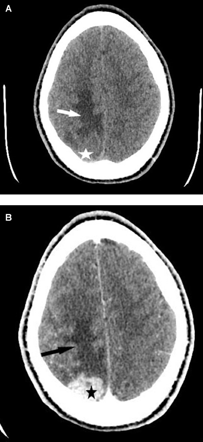
Meningeal tuberculoma is one of the most serious sites of tuberculosis. Its incidence varies depending on the geographical area, rare in Western countries and frequent in developing countries where it represents 5% to 10% of intracranial masses. We report the case of a 21-year-old male patient with no particular medical history from Africa and living in Europe for more than a year, is hospitalized for an isolated inaugural, generalized, afebrile seizure in whom the scanner and cerebral magnetic resonance imaging (MRI) revealed a meningeal mass with significant glove finger edema suggesting a primary brain tumor. Surgical excision and anatomopathological analysis of the excisional piece allowed the diagnosis of tuberculoma. Meningeal tuberculoma is a source of diagnostic error because its clinical and radiological expression can mimic a brain tumor. This is an etiology that should not be ignored in the face of a meningeal mass in any subject coming from or living in a region with a high endemic tuberculosis.
脑膜结核瘤是结核病最严重的发病部位之一。其发病率因地理区域而异,在西方国家罕见,在发展中国家较为常见,在这些国家它占颅内肿块的5%至10%。我们报告了一例21岁男性患者的病例,该患者来自非洲,无特殊病史,在欧洲生活一年多,因首次出现孤立的全身性无热惊厥住院,其脑部CT扫描和磁共振成像(MRI)显示有一个伴有明显指套样水肿的脑膜肿块,提示原发性脑肿瘤。对切除组织进行手术切除和解剖病理学分析后确诊为结核瘤。脑膜结核瘤是诊断错误的一个来源,因为其临床和影像学表现可酷似脑肿瘤。对于来自或生活在结核病高流行地区的任何出现脑膜肿块的患者,这种病因都不应被忽视。