Pargas Carlos, Franzone Jeanne M, Rogers Kenneth J, Artinian Frank, Santana Adolfredo, Shah Suken A, McGreal Cristina M, Kruse Richard W, Bober Michael B
Department of Orthopaedic Surgery, Nemours/Alfred I. duPont Hospital for Children, Wilmington, DE, USA.
Department of Orthopaedic Surgery, Akron Children's Hospital, Akron, OH, USA.
Bone Rep. 2020 Nov 21;13:100735. doi: 10.1016/j.bonr.2020.100735. eCollection 2020 Dec.
Osteogenesis imperfecta (OI) is a heterogeneous group of genetic disorders of connective tissue that cause skeletal fragility and extra-skeletal manifestations. Classically, four different types of OI were distinguished. Type 5 OI was added due to its distinct clinical and radiographic features. In 2012, two independent groups identified a recurrent heterozygous c.-14C>T mutation in as the responsible genetic change for this type of OI. To our knowledge, cervical kyphosis has not been identified in the literature as a finding in type 5 OI patients. This is a retrospective review of a cohort of patients with type 5 OI and a description of associated cervical spine deformity.
After institutional review board approval, a retrospective review identified 13 patients with type 5 OI. Clinical, radiologic, and genetic data from 2002 to 2020 were reviewed.
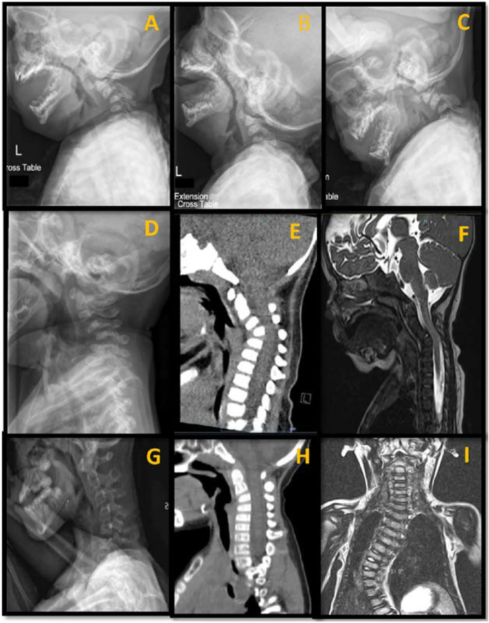
We identified 13 patients with clinical diagnosis of type 5 OI. Twelve had molecular confirmation and the classic , c.14C>T gene mutation was identified. The remaining individual did not undergo genetic testing. Dentinogenesis imperfecta was observed in one patient, while blue sclerae or hearing loss were not present. All patients had at least one fracture and four underwent intramedullary rodding. Radiologic features included subphyseal metaphyseal radiodense line in 12/13 patients (92%), interosseous membrane calcification in seven of 13 patients (54%) (more commonly noted in the upper extremities), and hypertrophic callus in six of 13 patients (46%). Thoracolumbar spinal deformities were seen in six of 13 patients (46%) with two of these individuals requiring surgery. Cervical kyphosis was noted in nine of 13 individuals (69%) ranging in age from 3 months to 22 years. Anterior wedging of the cervical vertebral bodies was noted in the absence of any fractures. Six of nine individuals demonstrated listhesis of C2-C3 or C3-C4 segment. Magnetic resonance imaging studies were performed and reviewed in patients with cervical kyphosis and subluxation; three patients showed narrowing of spinal canal without cervical cord compression and one asymptomatic patient showed impingement of the spinal cord.
Cervical kyphosis appears to be a common feature of type 5 OI. It can be a presenting and apparently life-long association and does not appear to be caused by vertebral body fractures. Evaluation for cervical kyphosis should be performed in patients with a suspected or confirmed diagnosis of type 5 OI. Furthermore, if cervical kyphosis is noted in an individual with OI, type 5 OI should be considered.Level of evidence: IV.
成骨不全(OI)是一组结缔组织遗传性疾病的异质性群体,可导致骨骼脆弱和骨骼外表现。传统上,OI分为四种不同类型。由于其独特的临床和影像学特征,增加了5型OI。2012年,两个独立的研究小组发现,作为这种类型OI的致病基因变化,在 中存在一个反复出现的杂合子c.-14C>T突变。据我们所知,文献中尚未将颈椎后凸确定为5型OI患者的一项发现。这是一项对一组5型OI患者的回顾性研究,并描述了相关的颈椎畸形。
经机构审查委员会批准后,进行回顾性研究,确定了13例5型OI患者。回顾了2002年至2020年的临床、放射学和基因数据。
我们确定了13例临床诊断为5型OI的患者。12例有分子学确诊,并鉴定出经典的 、c.14C>T基因突变。其余个体未进行基因检测。在1例患者中观察到牙本质发育不全,而未出现蓝色巩膜或听力丧失。所有患者至少有一处骨折,4例接受了髓内棒植入术。放射学特征包括12/13例患者(92%)出现骨骺下干骺端致密线,13例患者中有7例(54%)出现骨间膜钙化(在上肢更常见),13例患者中有6例(46%)出现肥大性骨痂。13例患者中有6例(46%)出现胸腰椎脊柱畸形,其中2例需要手术治疗。13例患者中有9例(69%)出现颈椎后凸,年龄从3个月至22岁不等。在没有任何骨折的情况下,发现颈椎椎体前缘楔形变。9例患者中有6例出现C2-C3或C3-C4节段滑脱。对颈椎后凸和半脱位患者进行了磁共振成像研究并进行了分析;3例患者显示椎管狭窄但无颈髓受压,1例无症状患者显示脊髓受压。
颈椎后凸似乎是5型OI的一个常见特征。它可能是一个呈现且明显终身存在的关联,似乎不是由椎体骨折引起的。对于疑似或确诊为5型OI的患者,应进行颈椎后凸评估。此外,如果在OI患者中发现颈椎后凸,应考虑5型OI。证据级别:IV级。