• 文献检索
  • 文档翻译
  • 深度研究
  • 学术资讯
  • Suppr Zotero 插件Zotero 插件
  • 邀请有礼
  • 套餐&价格
  • 历史记录
应用&插件
Suppr Zotero 插件Zotero 插件浏览器插件Mac 客户端Windows 客户端微信小程序
定价
高级版会员购买积分包购买API积分包
服务
文献检索文档翻译深度研究API 文档MCP 服务
关于我们
关于 Suppr公司介绍联系我们用户协议隐私条款
关注我们

Suppr 超能文献

核心技术专利:CN118964589B侵权必究
粤ICP备2023148730 号-1Suppr @ 2026

文献检索

告别复杂PubMed语法,用中文像聊天一样搜索,搜遍4000万医学文献。AI智能推荐,让科研检索更轻松。

立即免费搜索

文件翻译

保留排版,准确专业,支持PDF/Word/PPT等文件格式,支持 12+语言互译。

免费翻译文档

深度研究

AI帮你快速写综述,25分钟生成高质量综述,智能提取关键信息,辅助科研写作。

立即免费体验

从影像学角度诊断进展性多发性硬化症:综述。

Diagnosis of Progressive Multiple Sclerosis From the Imaging Perspective: A Review.

机构信息

Neuroimaging Research Unit, Institute of Experimental Neurology, Division of Neuroscience, Istituto di Ricovero e di Cura a Carattere Scientifico (IRCCS) San Raffaele Scientific Institute, Milan, Italy.

Neurology Unit, IRCCS San Raffaele Scientific Institute, Milan, Italy.

出版信息

JAMA Neurol. 2021 Mar 1;78(3):351-364. doi: 10.1001/jamaneurol.2020.4689.

DOI:10.1001/jamaneurol.2020.4689
PMID:33315071
原文链接:https://pmc.ncbi.nlm.nih.gov/articles/PMC11382596/
Abstract

IMPORTANCE

Although magnetic resonance imaging (MRI) is useful for monitoring disease dissemination in space and over time and excluding multiple sclerosis (MS) mimics, there has been less application of MRI to progressive MS, including diagnosing primary progressive (PP) MS and identifying patients with relapsing-remitting (RR) MS who are at risk of developing secondary progressive (SP) MS. This review addresses clinical application of MRI for both diagnosis and prognosis of progressive MS.

OBSERVATIONS

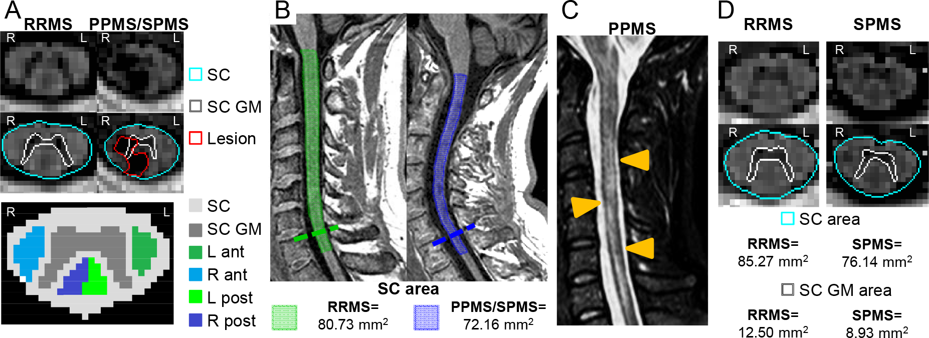
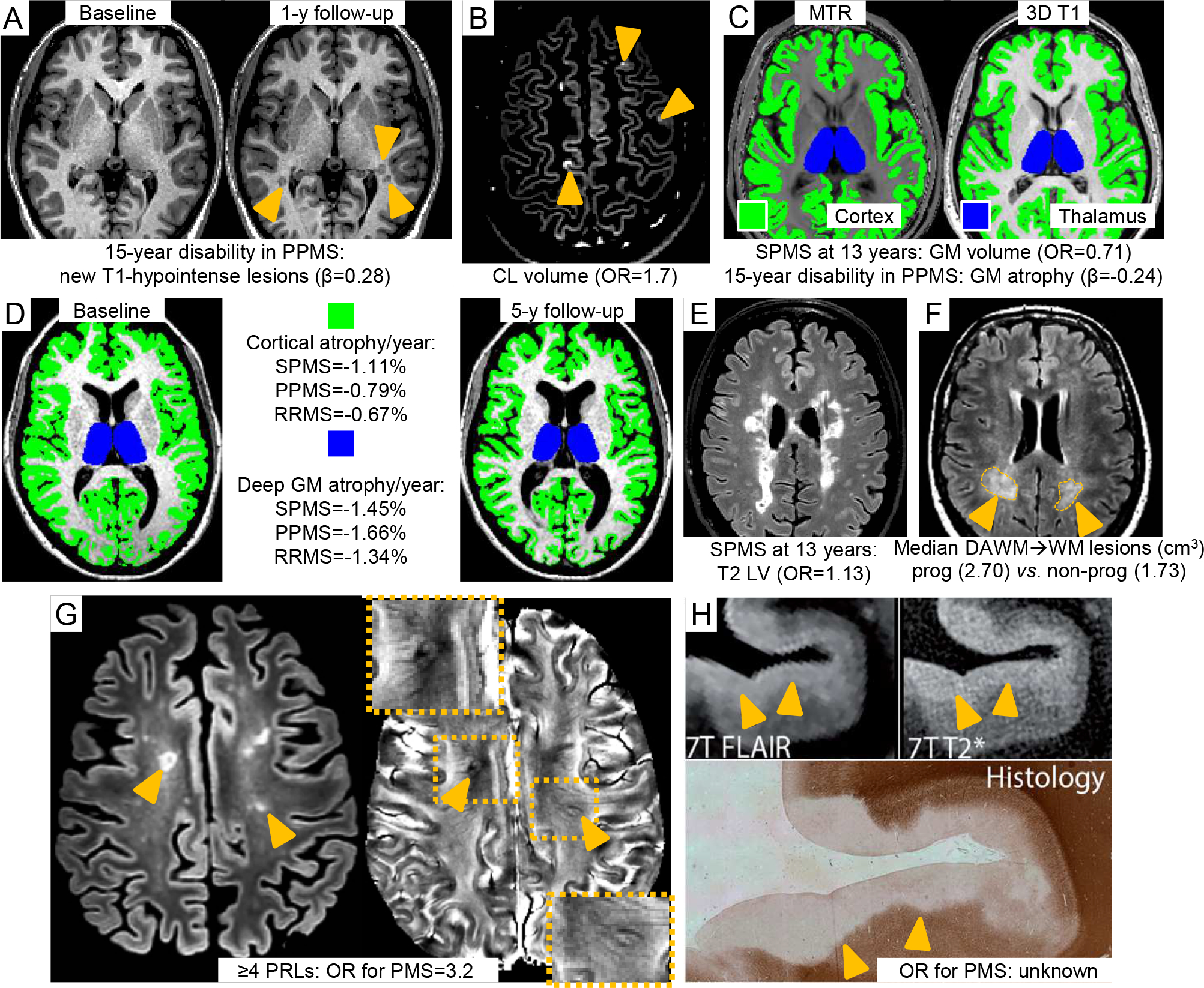
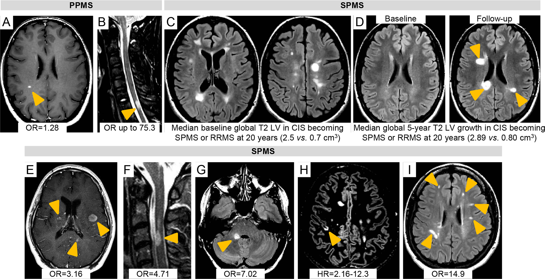
Although nonspecific, some spinal cord imaging features (diffuse abnormalities and lesions involving gray matter [GM] and ≥2 white matter columns) are typical of PPMS. In patients with PPMS and those with relapse-onset MS, location of lesions in critical central nervous system regions (spinal cord, infratentorial regions, and GM) and MRI-detected high inflammatory activity in the first years after diagnosis are risk factors for long-term disability and future progressive disease course. These measures are evaluable in clinical practice. In patients with established MS, GM involvement and neurodegeneration are associated with accelerated clinical worsening. Subpial demyelination and slowly expanding lesions are novel indicators of progressive MS.

CONCLUSIONS AND RELEVANCE

Diagnosis of PPMS is more challenging than diagnosis of RRMS. No qualitative clinical, immunological, histopathological, or neuroimaging features differentiate PPMS and SPMS; both are characterized by imaging findings reflecting neurodegeneration and are also impacted by aging and comorbidities. Unmet diagnostic needs include identification of MRI markers capable of distinguishing PPMS from RRMS and predicting the evolution of RRMS to SPMS. Integration of multiple parameters will likely be essential to achieve these aims.

摘要

重要性

尽管磁共振成像(MRI)可用于监测疾病在空间和时间上的扩散,并排除多发性硬化症(MS)的类似物,但在进展性 MS 中应用 MRI 的情况较少,包括诊断原发性进展型(PP)MS 并识别出有发展为继发性进展型(SP)MS 风险的缓解复发型(RR)MS 患者。本综述介绍了 MRI 在进展性 MS 的诊断和预后中的临床应用。

观察结果

尽管非特异性,但一些脊髓成像特征(弥漫性异常和累及灰质[GM]和≥2 个白质柱的病变)是 PPMS 的典型特征。在 PPMS 患者和发病后复发的 MS 患者中,病变位于关键中枢神经系统区域(脊髓、颅后窝区域和 GM)以及诊断后最初几年 MRI 检测到的高炎症活性是长期残疾和未来进行性疾病过程的风险因素。这些措施可在临床实践中评估。在已确诊的 MS 患者中,GM 受累和神经退行性变与加速临床恶化有关。软脑膜下脱髓鞘和缓慢扩展的病变是进展性 MS 的新指标。

结论和相关性

PPMS 的诊断比 RRMS 的诊断更具挑战性。没有定性的临床、免疫学、组织病理学或神经影像学特征可以区分 PPMS 和 SPMS;两者均以反映神经退行性变的影像学表现为特征,并且还受到衰老和合并症的影响。未满足的诊断需求包括识别能够将 PPMS 与 RRMS 区分开的 MRI 标志物,并预测 RRMS 向 SPMS 的演变。整合多个参数可能对于实现这些目标至关重要。

https://cdn.ncbi.nlm.nih.gov/pmc/blobs/15ef/11382596/a10c5c896d2b/nihms-2018594-f0003.jpg
https://cdn.ncbi.nlm.nih.gov/pmc/blobs/15ef/11382596/63a8cd84c2d5/nihms-2018594-f0001.jpg
https://cdn.ncbi.nlm.nih.gov/pmc/blobs/15ef/11382596/f7e0602d2888/nihms-2018594-f0002.jpg
https://cdn.ncbi.nlm.nih.gov/pmc/blobs/15ef/11382596/a10c5c896d2b/nihms-2018594-f0003.jpg
https://cdn.ncbi.nlm.nih.gov/pmc/blobs/15ef/11382596/63a8cd84c2d5/nihms-2018594-f0001.jpg
https://cdn.ncbi.nlm.nih.gov/pmc/blobs/15ef/11382596/f7e0602d2888/nihms-2018594-f0002.jpg
https://cdn.ncbi.nlm.nih.gov/pmc/blobs/15ef/11382596/a10c5c896d2b/nihms-2018594-f0003.jpg

相似文献

1
Diagnosis of Progressive Multiple Sclerosis From the Imaging Perspective: A Review.从影像学角度诊断进展性多发性硬化症:综述。
JAMA Neurol. 2021 Mar 1;78(3):351-364. doi: 10.1001/jamaneurol.2020.4689.
2
Grey matter involvement by focal cervical spinal cord lesions is associated with progressive multiple sclerosis.局灶性颈脊髓病变累及灰质与进展型多发性硬化症相关。
Mult Scler. 2016 Jun;22(7):910-20. doi: 10.1177/1352458515604905. Epub 2015 Oct 2.
3
Neurological disability and brain grey matter atrophy in primary progressive multiple sclerosis are determined by microstructural lesional changes, but not by lesion load.原发性进行性多发性硬化症中的神经功能障碍和脑灰质萎缩是由微观结构病变改变决定的,而非由病灶负荷决定。
J Neurol. 2025 Apr 1;272(4):302. doi: 10.1007/s00415-025-13043-x.
4
Magnetic Resonance Imaging in Primary Progressive Multiple Sclerosis Patients : Review.原发性进行性多发性硬化症患者的磁共振成像:综述。
Clin Neuroradiol. 2022 Sep;32(3):625-641. doi: 10.1007/s00062-022-01144-3. Epub 2022 Mar 8.
5
Preferential spinal cord volume loss in primary progressive multiple sclerosis.原发性进行性多发性硬化症中脊髓体积优先损失。
Mult Scler. 2019 Jun;25(7):947-957. doi: 10.1177/1352458518775006. Epub 2018 May 21.
6
Pathogenic Mechanisms Associated With Different Clinical Courses of Multiple Sclerosis.与多发性硬化症不同临床病程相关的发病机制。
Front Immunol. 2019 Jan 10;9:3116. doi: 10.3389/fimmu.2018.03116. eCollection 2018.
7
Multiple sclerosis by phenotype in Germany.德国基于表型的多发性硬化症。
Mult Scler Relat Disord. 2022 Jan;57:103326. doi: 10.1016/j.msard.2021.103326. Epub 2021 Oct 10.
8
Cervical cord area is associated with infratentorial grey and white matter volume predominantly in relapsing-remitting multiple sclerosis: A study using semi-automated cord volumetry and voxel-based morphometry.在复发缓解型多发性硬化症中,颈髓区域主要与幕下灰质和白质体积相关:一项使用半自动脊髓容积测量法和基于体素的形态测量学的研究
Mult Scler Relat Disord. 2015 May;4(3):264-72. doi: 10.1016/j.msard.2015.04.003. Epub 2015 Apr 15.
9
Critical spinal cord lesions associate with secondary progressive motor impairment in long-standing MS: A population-based case-control study.长期多发性硬化症患者中严重的脊髓病变与继发性进行性运动功能障碍相关:一项基于人群的病例对照研究。
Mult Scler. 2021 Apr;27(5):667-673. doi: 10.1177/1352458520929192. Epub 2020 Jun 18.
10
7 T imaging reveals a gradient in spinal cord lesion distribution in multiple sclerosis.7T 成像显示多发性硬化症的脊髓病变分布呈梯度变化。
Brain. 2020 Oct 1;143(10):2973-2987. doi: 10.1093/brain/awaa249.

引用本文的文献

1
AI-driven reclassification of multiple sclerosis progression.人工智能驱动的多发性硬化症病情进展重新分类
Nat Med. 2025 Aug 20. doi: 10.1038/s41591-025-03901-6.
2
The use of high-efficacy disease-modifying therapies in multiple sclerosis: recommendations from an expert Delphi consensus.多发性硬化症高效疾病修正疗法的应用:专家德尔菲共识推荐意见
J Neurol. 2025 Aug 10;272(9):565. doi: 10.1007/s00415-025-13293-9.
3
The Role of MRI Lesions in Identifying Secondary Progressive Multiple Sclerosis: A Comprehensive Review.MRI病变在识别继发进展型多发性硬化中的作用:一项综述

本文引用的文献

1
Progressive Multiple Sclerosis Is Associated with Faster and Specific Retinal Layer Atrophy.进行性多发性硬化症与更快和更特定的视网膜层萎缩有关。
Ann Neurol. 2020 Jun;87(6):885-896. doi: 10.1002/ana.25738. Epub 2020 Apr 28.
2
Conversion of diffusely abnormal white matter to focal lesions is linked to progression in secondary progressive multiple sclerosis.弥漫性异常白质向局灶性病变的转变与继发进展型多发性硬化症的病情进展相关。
Mult Scler. 2021 Feb;27(2):208-219. doi: 10.1177/1352458520912172. Epub 2020 Mar 23.
3
Volume loss in the deep gray matter and thalamic subnuclei: a longitudinal study on disability progression in multiple sclerosis.
J Clin Med. 2025 Jun 10;14(12):4114. doi: 10.3390/jcm14124114.
4
Diffusion- and Tractography-Based Characterization of Tissue Damage Within and Surrounding Paramagnetic Rim Lesions in Multiple Sclerosis.基于扩散和纤维束成像的多发性硬化症顺磁性边缘病变内部及周围组织损伤特征分析
AJNR Am J Neuroradiol. 2025 Mar 4;46(3):611-619. doi: 10.3174/ajnr.A8524.
5
MRI versus relapse: optimal activity monitoring for management of progressive multiple sclerosis.磁共振成像与复发:用于进行性多发性硬化症管理的最佳活动监测
Brain Commun. 2025 Jan 20;7(1):fcaf010. doi: 10.1093/braincomms/fcaf010. eCollection 2025.
6
Upper cervical spinal cord atrophy in MS: Sex, menopause, and neurodegeneration.多发性硬化症中上颈段脊髓萎缩:性别、绝经与神经退行性变
Mult Scler. 2025 Mar;31(3):278-289. doi: 10.1177/13524585241311441. Epub 2025 Jan 18.
7
Enhancing classification of active and non-active lesions in multiple sclerosis: machine learning models and feature selection techniques.增强多发性硬化症中活动性和非活动性病变的分类:机器学习模型与特征选择技术
BMC Med Imaging. 2024 Dec 20;24(1):345. doi: 10.1186/s12880-024-01528-6.
8
Towards a Unified Set of Diagnostic Criteria for Multiple Sclerosis.迈向多发性硬化症统一诊断标准集
Ann Neurol. 2025 Mar;97(3):571-582. doi: 10.1002/ana.27145. Epub 2024 Nov 28.
9
Vitamin D receptor gene polymorphisms in patients with relapsing multiple sclerosis.复发型多发性硬化症患者维生素D受体基因多态性
Eur J Transl Myol. 2025 Mar 31;35(1). doi: 10.4081/ejtm.2024.12993. Epub 2024 Nov 21.
10
Early regional cerebral grey matter damage predicts long-term cognitive impairment phenotypes in multiple sclerosis: a 20-year study.早期脑区灰质损伤可预测多发性硬化症的长期认知障碍表型:一项20年的研究。
Brain Commun. 2024 Oct 12;6(6):fcae355. doi: 10.1093/braincomms/fcae355. eCollection 2024.
脑深部灰质和丘脑亚核体积损失:多发性硬化残疾进展的纵向研究。
J Neurol. 2020 May;267(5):1536-1546. doi: 10.1007/s00415-020-09740-4. Epub 2020 Feb 10.
4
7T MRI cerebral leptomeningeal enhancement is common in relapsing-remitting multiple sclerosis and is associated with cortical and thalamic lesions.7T MRI 脑软脑膜强化在复发缓解型多发性硬化症中很常见,与皮质和丘脑病变有关。
Mult Scler. 2020 Feb;26(2):177-187. doi: 10.1177/1352458519885106. Epub 2019 Nov 12.
5
A 30-Year Clinical and Magnetic Resonance Imaging Observational Study of Multiple Sclerosis and Clinically Isolated Syndromes.一项多发性硬化症和临床孤立综合征的 30 年临床和磁共振成像观察研究。
Ann Neurol. 2020 Jan;87(1):63-74. doi: 10.1002/ana.25637. Epub 2019 Nov 22.
6
Clinically relevant cranio-caudal patterns of cervical cord atrophy evolution in MS.MS 中颈髓萎缩演变的临床相关头尾模式。
Neurology. 2019 Nov 12;93(20):e1852-e1866. doi: 10.1212/WNL.0000000000008466. Epub 2019 Oct 14.
7
Serum neurofilament light chain level associations with clinical and cognitive performance in multiple sclerosis: A longitudinal retrospective 5-year study.血清神经丝轻链水平与多发性硬化症的临床和认知表现的相关性:一项纵向回顾性 5 年研究。
Mult Scler. 2020 Nov;26(13):1670-1681. doi: 10.1177/1352458519881428. Epub 2019 Oct 15.
8
Chronic white matter lesion activity predicts clinical progression in primary progressive multiple sclerosis.慢性白质病变活动可预测原发性进行性多发性硬化症的临床进展。
Brain. 2019 Sep 1;142(9):2787-2799. doi: 10.1093/brain/awz212.
9
Association of Chronic Active Multiple Sclerosis Lesions With Disability In Vivo.慢性活动性多发性硬化症病灶与体内残疾的关联。
JAMA Neurol. 2019 Dec 1;76(12):1474-1483. doi: 10.1001/jamaneurol.2019.2399.
10
Association Between Serum Neurofilament Light Chain Levels and Long-term Disease Course Among Patients With Multiple Sclerosis Followed up for 12 Years.血清神经丝轻链水平与随访12年的多发性硬化症患者长期病程之间的关联
JAMA Neurol. 2019 Nov 1;76(11):1359-1366. doi: 10.1001/jamaneurol.2019.2137.