Department of Immunology, School of Basic Medical Sciences, Peking University, Beijing 100191, China.
NHC Key Laboratory of Medical Immunology, Peking University, Beijing 100191, China.
Int J Mol Sci. 2020 Dec 9;21(24):9379. doi: 10.3390/ijms21249379.
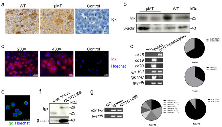
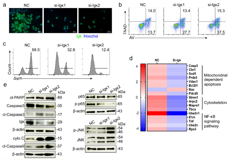
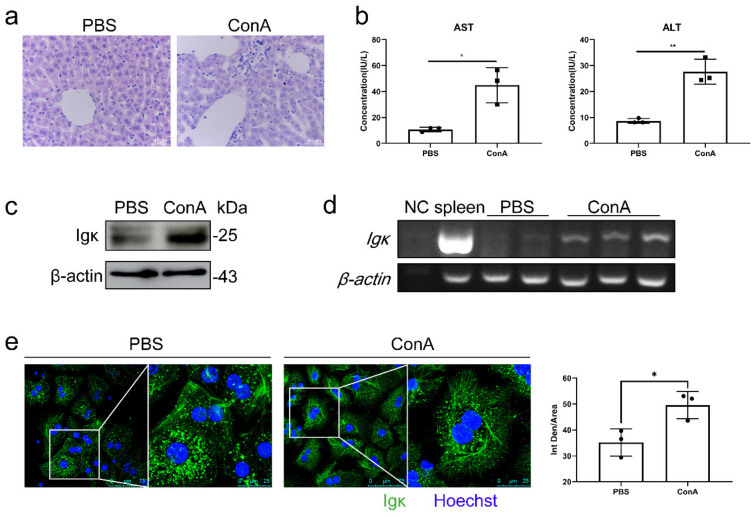
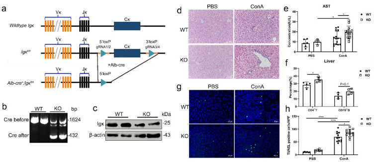
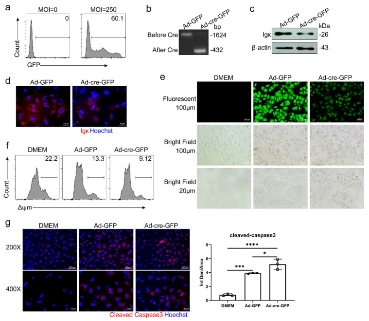
Immunoglobulin (Igκ) has been reported to be expressed in sorted liver epithelial cells of μMT mice, and the sequence characteristics of hepatocyte-derived Igκ were different from those of classical B-cell-derived Igκ. However, the physiological function of hepatocyte-derived Igκ is still unclear. The expression of Igκ was firstly identified in primary hepatocytes and normal liver cell line (NCTC1469), and hepatocyte-derived Igκ expression was elevated and displayed unique localization in hepatocytes of concanavalin A (ConA)-induced hepatitis model. Moreover, knockout mice were more sensitive to ConA-induced hepatitis and had higher serum aspartate aminotransferase (AST) levels, more severe histological injury and a greater number of terminal deoxynucleotide transferase-mediated deoxyuridine triphosphate nick end-labeling (TUNEL)-positive cells as compared with littermate controls. Furthermore, knockdown of in primary hepatocytes and NCTC1469 cells led to accelerated activation of the mitochondrial death pathway and caspase-3 cleavage in vitro, which might be related to inhibition of NF-κB signaling pathway and activation of JNK via the cytoskeleton dynamics. Taken together, these results indicate that hepatocyte-derived Igκ mediates cellular resistance to ConA-induced liver injury by inhibiting activation of caspase-3 and the mitochondrial death pathway, suggesting that Igκ plays an important role in hepatocyte survival and exerts a protective effect against ConA-induced liver injury in mice.
免疫球蛋白(Igκ)已被报道在 μMT 小鼠的分离肝上皮细胞中表达,并且肝细胞来源的 Igκ 的序列特征与经典 B 细胞来源的 Igκ 不同。然而,肝细胞来源的 Igκ 的生理功能尚不清楚。Igκ 的表达首先在原代肝细胞和正常肝细胞系(NCTC1469)中被鉴定,并且在伴刀豆球蛋白 A(ConA)诱导的肝炎模型的肝细胞中,肝细胞来源的 Igκ 表达上调并呈现独特的定位。此外,与同窝对照相比,敲除小鼠对 ConA 诱导的肝炎更敏感,血清天冬氨酸氨基转移酶(AST)水平更高,组织学损伤更严重,末端脱氧核苷酸转移酶介导的脱氧尿苷三磷酸缺口末端标记(TUNEL)阳性细胞数量更多。此外,在原代肝细胞和 NCTC1469 细胞中敲低 会导致体外线粒体死亡途径的激活和 caspase-3 切割加速,这可能与抑制 NF-κB 信号通路和通过细胞骨架动力学激活 JNK 有关。总之,这些结果表明,肝细胞来源的 Igκ 通过抑制 caspase-3 和线粒体死亡途径的激活来介导细胞对 ConA 诱导的肝损伤的抵抗,表明 Igκ 在肝细胞存活中发挥重要作用,并对 ConA 诱导的小鼠肝损伤发挥保护作用。