Aggeli Constantina, Dimitroglou Yannis, Raftopoulos Leonidas, Sarri Georgia, Mavrogeni Sophie, Wong Joyce, Tsiamis Eleftherios, Tsioufis Costas
First Department of Cardiology, General Hospital of Athens Hippokration, University of Athens Medical School, 11527 Athens, Attica, Greece.
Department of Cardiology, Onassis Cardiac Surgery Centre, 17674 Kallithea, Attica, Greece.
Diagnostics (Basel). 2020 Dec 14;10(12):1088. doi: 10.3390/diagnostics10121088.
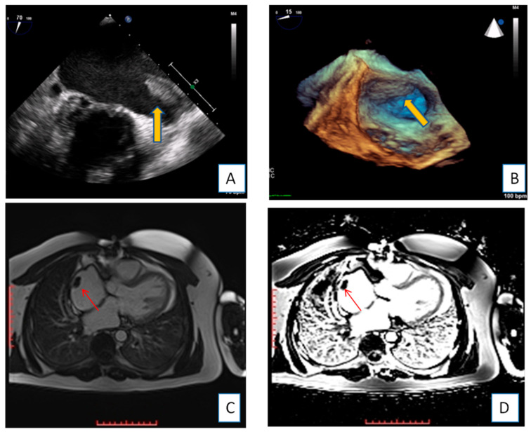
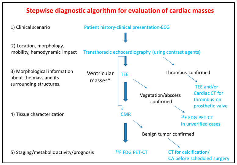
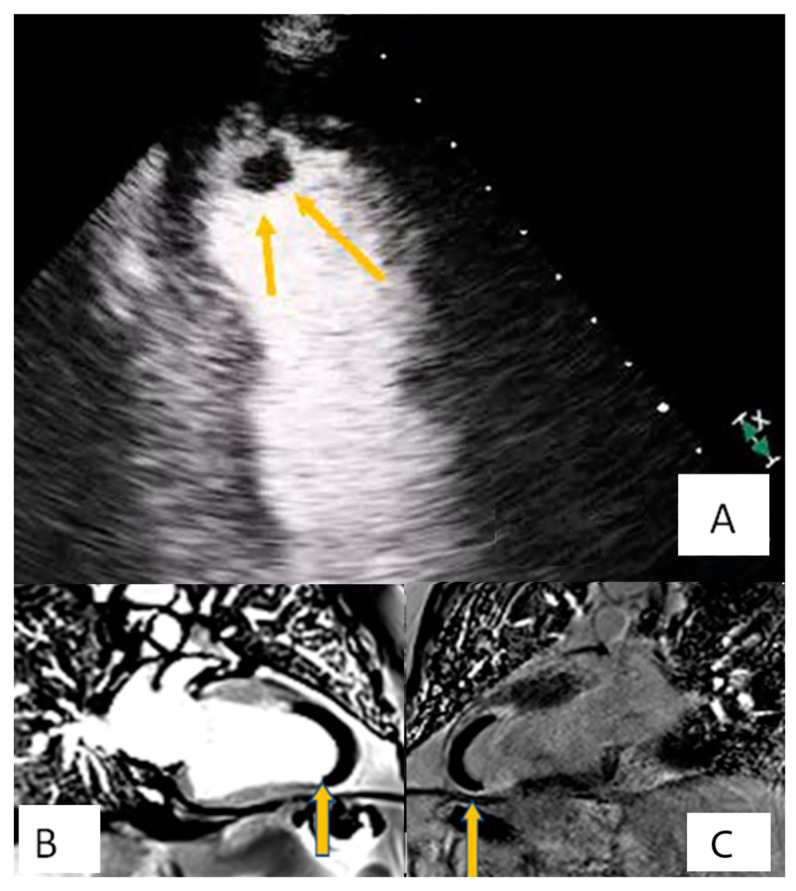
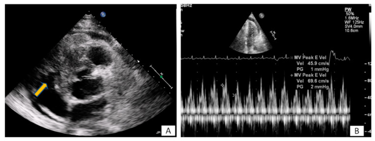
Cardiac masses are space occupying lesions within the cardiac cavities or adjacent to the pericardium. They include frequently diagnosed clinical entities such as clots and vegetations, common benign tumors such as myxomas and papillary fibroelastomas and uncommon benign or malignant primary or metastatic tumors. Given their diversity, there are no guidelines or consensus statements regarding the best diagnostic or therapeutic approach. In the past, diagnosis used to be made by the histological specimens after surgery or during the post-mortem examination. Nevertheless, evolution and increased availability of cardiovascular imaging modalities has enabled better characterization of the masses and the surrounding tissue. Transthoracic echocardiography using contrast agents can evaluate the location, the morphology and the perfusion of the mass as well as its hemodynamic effect. Transesophageal echocardiography has increased spatial and temporal resolution; hence it is superior in depicting small highly mobile masses. Cardiac magnetic resonance and cardiac computed tomography are complementary providing tissue characterization. The scope of this review is to present the role of cardiovascular imaging in the differential diagnosis of cardiac masses and to propose a step-wise diagnostic algorithm, taking into account the epidemiology and clinical presentation of the cardiac masses, as well as the availability and the incremental value of each imaging modality.
心脏肿物是指位于心腔内或心包附近的占位性病变。它们包括常见的临床诊断实体,如血栓和赘生物;常见的良性肿瘤,如黏液瘤和乳头状纤维弹性瘤;以及罕见的良性或恶性原发性或转移性肿瘤。鉴于其多样性,目前尚无关于最佳诊断或治疗方法的指南或共识声明。过去,诊断通常通过手术切除后的组织学标本或尸检来进行。然而,心血管成像技术的发展和可用性的提高,使得对肿物及其周围组织的特征有了更好的描述。使用造影剂的经胸超声心动图可以评估肿物的位置、形态、灌注情况及其血流动力学效应。经食管超声心动图具有更高的空间和时间分辨率,因此在显示小的高活动性肿物方面更具优势。心脏磁共振成像和心脏计算机断层扫描具有互补性,可提供组织特征。本综述的目的是介绍心血管成像在心脏肿物鉴别诊断中的作用,并提出一种逐步诊断算法,同时考虑心脏肿物的流行病学和临床表现,以及每种成像方式的可用性和增加值。