Hannikainen Paavali A, Kosa Peter, Barbour Christopher, Bielekova Bibiana
Neuroimmunological Diseases Section, Laboratory of Clinical Immunology and Microbiology, National Institute of Allergy and Infectious Diseases, National Institutes of Health, Bethesda, MD, United States.
Front Neurol. 2020 Nov 27;11:565957. doi: 10.3389/fneur.2020.565957. eCollection 2020.
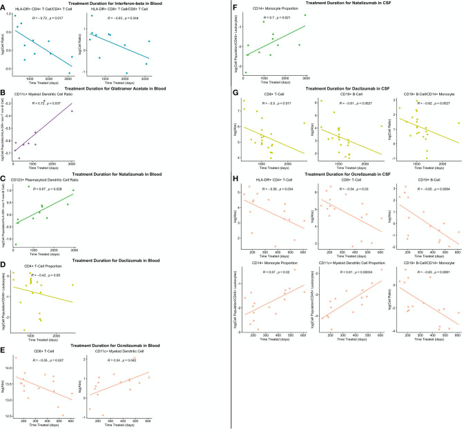
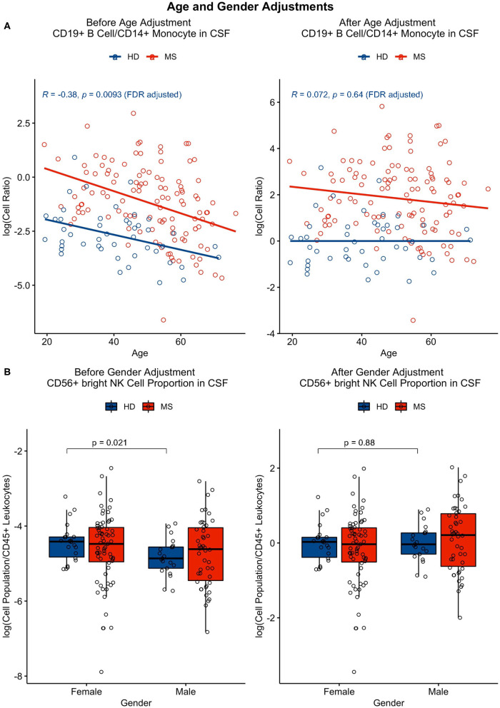
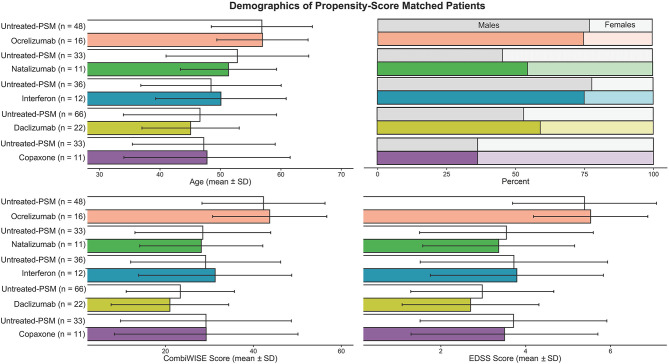
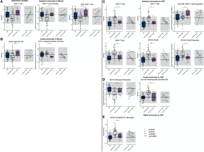
Quantifying cell subpopulations in biological fluids aids in diagnosis and understanding of the mechanisms of injury. Although much has been learned from cerebrospinal fluid (CSF) flow cytometry in neuroimmunological disorders, such as multiple sclerosis (MS), previous studies did not contain enough healthy donors (HD) to derive age- and gender-related normative data and sufficient heterogeneity of other inflammatory neurological disease (OIND) controls to identify MS specific changes. The goals of this blinded training and validation study of MS patients and embedded controls, representing 1,240 prospectively acquired paired CSF/blood samples from 588 subjects was (1) to define physiological age-/gender-related changes in CSF cells, (2) to define/validate cellular abnormalities in blood and CSF of untreated MS through disease duration (DD) and determine which are MS-specific, and (3) to compare effect(s) of low-efficacy (i.e., interferon-beta [IFN-beta] and glatiramer acetate [GA]) and high-efficacy drugs (i.e., natalizumab, daclizumab, and ocrelizumab) on MS-related cellular abnormalities using propensity score matching. Physiological gender differences are less pronounced in the CSF compared to blood, and age-related changes suggest decreased immunosurveillance of CNS by activated HLA-DR+T cells associated with natural aging. Results from patient samples support the concept of MS being immunologically single disease evolving in time. Initially, peripherally activated innate and adaptive immune cells migrate into CSF to form MS lesions. With progression, T cells (CD8+ > CD4+), NK cells, and myeloid dendritic cells are depleted from blood as they continue to accumulate, together with B cells, in the CSF and migrate to CNS tissue, forming compartmentalized inflammation. All MS drugs inhibit non-physiological accumulation of immune cells in the CSF. Although low-efficacy drugs tend to normalize it, high-efficacy drugs overshoot some aspects of CSF physiology, suggesting impairment of CNS immunosurveillance. Comparable inhibition of MS-related CSF abnormalities advocates changes within CNS parenchyma responsible for differences in drug efficacy on MS disability progression. Video summarizing all results may become useful educational tool.
对生物体液中的细胞亚群进行定量有助于疾病诊断和了解损伤机制。尽管通过脑脊液(CSF)流式细胞术在神经免疫疾病(如多发性硬化症(MS))方面已取得了很多认识,但先前的研究没有纳入足够多的健康供体(HD)来得出与年龄和性别相关的规范数据,也没有纳入足够异质性的其他炎性神经系统疾病(OIND)对照来识别MS的特异性变化。这项针对MS患者及嵌入式对照的双盲训练和验证研究,涵盖了来自588名受试者的1240份前瞻性采集的配对CSF/血液样本,其目标是:(1)确定CSF细胞中与生理年龄/性别相关的变化;(2)通过疾病持续时间(DD)定义/验证未经治疗的MS患者血液和CSF中的细胞异常,并确定哪些是MS特异性的;(3)使用倾向得分匹配比较低效药物(即干扰素-β[IFN-β]和醋酸格拉替雷[GA])和高效药物(即那他珠单抗、达利珠单抗和奥瑞珠单抗)对MS相关细胞异常的影响。与血液相比,CSF中的生理性性别差异不太明显,与年龄相关的变化表明,随着自然衰老,活化的HLA-DR⁺T细胞对中枢神经系统的免疫监视作用减弱。患者样本的结果支持MS是一种随时间演变的免疫单一疾病的概念。最初,外周活化的先天性和适应性免疫细胞迁移到CSF中形成MS病变。随着疾病进展,T细胞(CD8⁺>CD4⁺)、NK细胞和髓样树突状细胞从血液中耗竭,因为它们继续与B细胞一起在CSF中积聚并迁移到中枢神经系统组织,形成分隔性炎症。所有MS药物都抑制免疫细胞在CSF中的非生理性积聚。尽管低效药物倾向于使其正常化,但高效药物在某些方面超过了CSF生理学水平,提示中枢神经系统免疫监视受损。对MS相关CSF异常的类似抑制表明,中枢神经系统实质内的变化是导致药物对MS残疾进展疗效差异的原因。总结所有结果的视频可能会成为有用的教育工具。