Yousaf Amman, Tayyab Ahmad, Anil Muhammad Sana Ullah, Ahmed Mohamed Mohamed Helmi, Ahmed Sana Sayed Hussein Badr Ahmed, Alobadli Amal
Radiology, Hamad General Hospital, Doha, QAT.
Radiology, Services Institute of Medical Sciences, Lahore, PAK.
Cureus. 2020 Nov 14;12(11):e11480. doi: 10.7759/cureus.11480.
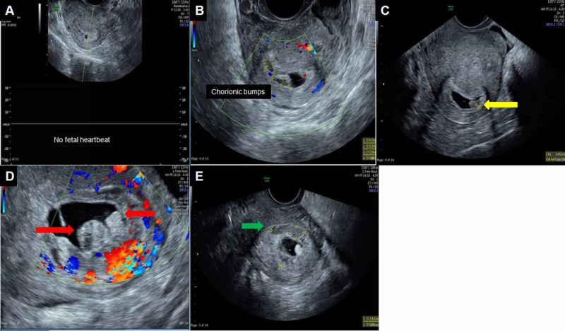
Background Chorionic bump is a rare condition defined as a bulge or protrusion from the choriodecidual surface into the gestational sac. The limited literature on this infrequent entity suggests that the pregnancies with multiple chorionic bumps mostly result in fetal demise. Aims To review the available literature and the patients from our institute having sonographic findings of chorionic bump and making the sonographers and radiologists aware of this known cause of first-trimester pregnancy loss. Study design A retrospective review of the cases diagnosed at our institute during the last four years. Methods and materials Single-center institutional data for four years (January 2016-December 2019) was accessed using ICD codes. IRB approval was waived owing to the anonymized use of patient data. Results Six female patients diagnosed with chorionic bump were included, with a mean age of 29.83±12 years. The average gestational age at the time of diagnosis was 8.16±3 weeks. The most common sonographic findings were a protrusion from the chorionic wall into the gestational sac cavity, having a central hypoechoic region with peripheral hyperechoic rim (isoechoic to the chorion) and having no vascularity (n=5), and the less common finding was a hyperechoic protrusion with no vascularity (n=1). n=5 had a single lesion, and n=1 had two lesions. The average diameter of the lesion in the largest dimension was 18±11 mm. n=3 pregnancies resulted in a first-trimester miscarriage, and n=3 pregnancies delivered healthy babies at term. Conclusions A chorionic bump significantly increases the risk of a first-trimester miscarriage.
绒毛膜隆起是一种罕见的情况,定义为从绒毛蜕膜表面向妊娠囊内的隆起或突出。关于这种罕见实体的文献有限,提示多绒毛膜隆起的妊娠大多导致胎儿死亡。目的:回顾现有文献以及我院有绒毛膜隆起超声表现的患者情况,使超声检查人员和放射科医生了解这种已知的孕早期流产原因。研究设计:对我院过去四年诊断的病例进行回顾性研究。方法和材料:使用国际疾病分类代码获取四年(2016年1月至2019年12月)的单中心机构数据。由于对患者数据进行匿名使用,因此无需获得机构审查委员会的批准。结果:纳入6例诊断为绒毛膜隆起的女性患者,平均年龄29.83±12岁。诊断时的平均孕周为8.16±3周。最常见的超声表现为从绒毛膜壁向妊娠囊腔内突出,中央为低回声区,周边为高回声边缘(与绒毛膜等回声)且无血管(n = 5),较不常见的表现为无血管的高回声突出(n = 1)。5例有单个病灶,1例有两个病灶。病灶最大径的平均直径为18±11mm。3例妊娠导致孕早期流产,3例妊娠足月分娩健康婴儿。结论:绒毛膜隆起显著增加孕早期流产的风险。