• 文献检索
  • 文档翻译
  • 深度研究
  • 学术资讯
  • Suppr Zotero 插件Zotero 插件
  • 邀请有礼
  • 套餐&价格
  • 历史记录
应用&插件
Suppr Zotero 插件Zotero 插件浏览器插件Mac 客户端Windows 客户端微信小程序
定价
高级版会员购买积分包购买API积分包
服务
文献检索文档翻译深度研究API 文档MCP 服务
关于我们
关于 Suppr公司介绍联系我们用户协议隐私条款
关注我们

Suppr 超能文献

核心技术专利:CN118964589B侵权必究
粤ICP备2023148730 号-1Suppr @ 2026

文献检索

告别复杂PubMed语法,用中文像聊天一样搜索,搜遍4000万医学文献。AI智能推荐,让科研检索更轻松。

立即免费搜索

文件翻译

保留排版,准确专业,支持PDF/Word/PPT等文件格式,支持 12+语言互译。

免费翻译文档

深度研究

AI帮你快速写综述,25分钟生成高质量综述,智能提取关键信息,辅助科研写作。

立即免费体验

影像主瘤表现对临床 IA 期(cIA)肺腺癌病理 N0(pN0)的选择。

Selection of Pathological N0 (pN0) in Clinical IA (cIA) Lung Adenocarcinoma by Imaging Findings of the Main Tumor.

机构信息

Department of Surgery, Teikyo University Hospital, Tokyo, Japan.

Department of Thoracic surgery, Aichi Cancer Center Hospital, Nagoya, Aichi, Japan.

出版信息

Ann Thorac Cardiovasc Surg. 2021 Aug 20;27(4):230-236. doi: 10.5761/atcs.oa.20-00240. Epub 2020 Dec 18.

DOI:10.5761/atcs.oa.20-00240
PMID:33342931
原文链接:https://pmc.ncbi.nlm.nih.gov/articles/PMC8374089/
Abstract

OBJECTIVES

We would like to clarify the imaging findings of the main tumor that may omit the requirement for lymph node dissection in clinical IA (cIA) lung adenocarcinoma.

METHODS

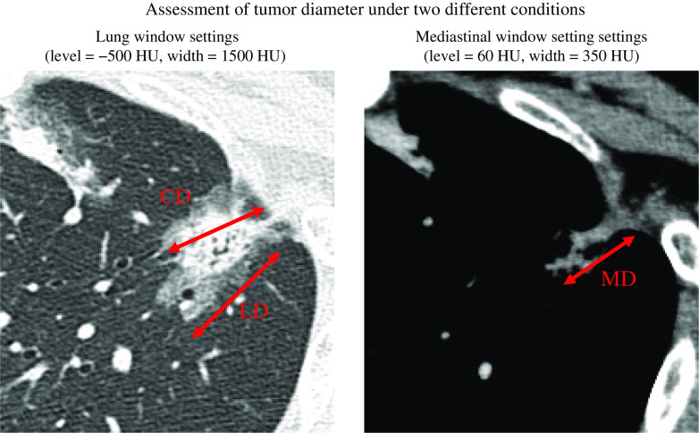
A total of 336 patients with cIA lung adenocarcinomas with normal preoperative carcinoembryonic antigen (CEA) who underwent surgical resection were analyzed. We investigated the association between various computed tomography (CT) imaging findings or the maximum standardized uptake value (SUVmax) of fluorodeoxyglucose-position emission tomography (FDG-PET) and lymph node metastasis. The maximum tumor diameter was calculated from the CT images using both the lung window setting (LD) and mediastinal window setting (MD). The diameter of the solid component (CD) was defined as consolidation diameter in lung window setting. The solid component ratio (C/T) was defined as CD/LD.

RESULTS

SUVmax, MD, and C/T were independent factors related to lymph node metastasis, but CD was not (p = 0.38). The conditions required for the positive predictive value (PPV) to reach 100% were 10.6 mm for MD, 12.5 mm for CD, and 0.55 for C/T. SUVmax did not reach 100%.

CONCLUSIONS

In cIA lung adenocarcinoma with CEA in the normal range, we found that it may be possible for lymph node dissection to be omitted by MD, CD, and C/T.

摘要

目的

我们希望阐明主要肿瘤的影像学表现,这些表现可能使临床 IA(cIA)肺腺癌患者免除淋巴结清扫的要求。

方法

分析了 336 例术前癌胚抗原(CEA)正常的 cIA 肺腺癌患者的资料。我们研究了各种 CT 成像表现或氟代脱氧葡萄糖正电子发射断层扫描(FDG-PET)的最大标准化摄取值(SUVmax)与淋巴结转移之间的关系。使用肺窗(LD)和纵隔窗(MD)从 CT 图像计算最大肿瘤直径。在 LD 中定义实性成分的直径为实变直径。实性成分比(C/T)定义为 CD/LD。

结果

SUVmax、MD 和 C/T 是与淋巴结转移相关的独立因素,但 CD 不是(p=0.38)。达到 100%阳性预测值(PPV)的条件分别为 MD 为 10.6mm、CD 为 12.5mm 和 C/T 为 0.55。SUVmax 未达到 100%。

结论

在 CEA 正常范围的 cIA 肺腺癌中,我们发现 MD、CD 和 C/T 可能使淋巴结清扫成为可能。

https://cdn.ncbi.nlm.nih.gov/pmc/blobs/a5bd/8374089/e84cae6c4b3a/atcs-27-230-g001.jpg

相似文献

1
Selection of Pathological N0 (pN0) in Clinical IA (cIA) Lung Adenocarcinoma by Imaging Findings of the Main Tumor.影像主瘤表现对临床 IA 期(cIA)肺腺癌病理 N0(pN0)的选择。
Ann Thorac Cardiovasc Surg. 2021 Aug 20;27(4):230-236. doi: 10.5761/atcs.oa.20-00240. Epub 2020 Dec 18.
2
Minor components of micropapillary and solid subtypes in lung invasive adenocarcinoma (≤ 3 cm): PET/CT findings and correlations with lymph node metastasis.肺浸润性腺癌(≤3cm)中小乳头状和实体型的次要成分:PET/CT 表现及其与淋巴结转移的相关性。
Radiol Med. 2020 Mar;125(3):257-264. doi: 10.1007/s11547-019-01112-x. Epub 2019 Dec 10.
3
Risk factors for lymph node metastasis and surgical methods in patients with early-stage peripheral lung adenocarcinoma presenting as ground glass opacity.表现为磨玻璃影的早期周围型肺腺癌患者淋巴结转移的危险因素及手术方法
J Cardiothorac Surg. 2020 Aug 12;15(1):121. doi: 10.1186/s13019-020-01167-2.
4
Prediction of pathologic node-negative clinical stage IA lung adenocarcinoma for optimal candidates undergoing sublobar resection.预测行亚肺叶切除术的病理淋巴结阴性ⅠA 期肺腺癌的最佳候选者。
J Thorac Cardiovasc Surg. 2012 Dec;144(6):1365-71. doi: 10.1016/j.jtcvs.2012.07.012. Epub 2012 Aug 9.
5
Factors associated with lymph node metastasis upstage after resection for patients with micropapillary lung adenocarcinoma.微乳头型肺腺癌患者术后淋巴结转移升级的相关因素。
Thorac Cancer. 2022 Jan;13(1):48-53. doi: 10.1111/1759-7714.14220. Epub 2021 Dec 5.
6
Long-Term Outcomes After Sublobar Resection Versus Lobectomy in Patients With Clinical Stage IA Lung Adenocarcinoma Meeting the Node-Negative Criteria Defined by High-Resolution Computed Tomography and [F]-Fluoro-2-Deoxy-d-Glucose Positron Emission Tomography.亚肺叶切除术与肺叶切除术治疗符合高分辨率计算机断层扫描和[F]-氟-2-脱氧-d-葡萄糖正电子发射断层扫描定义的淋巴结阴性标准的ⅠA 期肺腺癌患者的长期结果。
Clin Lung Cancer. 2021 May;22(3):e431-e437. doi: 10.1016/j.cllc.2020.06.013. Epub 2020 Jun 19.
7
Preoperative risk factors of lymph node metastasis in clinical N0 lung adenocarcinoma of 3 cm or less in diameter.直径 3cm 或以下临床 N0 肺腺癌的淋巴结转移术前危险因素。
BMC Surg. 2022 Apr 29;22(1):153. doi: 10.1186/s12893-022-01605-z.
8
Tumor-to-liver standard uptake ratio using fluorine-18 fluorodeoxyglucose positron emission tomography computed tomography effectively predict occult lymph node metastasis of non-small cell lung cancer patients.氟-18 氟代脱氧葡萄糖正电子发射断层扫描计算机断层扫描肿瘤与肝脏标准摄取比值可有效预测非小细胞肺癌患者隐匿性淋巴结转移。
Nucl Med Commun. 2020 May;41(5):459-468. doi: 10.1097/MNM.0000000000001173.
9
The maximum standardized uptake value of fluorodeoxyglucose positron emission tomography of the primary tumour is a good predictor of pathological nodal involvement in clinical N0 non-small-cell lung cancer.氟脱氧葡萄糖正电子发射断层扫描原发性肿瘤的最大标准化摄取值是临床 N0 非小细胞肺癌病理性淋巴结受累的良好预测指标。
Eur J Cardiothorac Surg. 2013 Jul;44(1):83-7. doi: 10.1093/ejcts/ezs604. Epub 2012 Dec 11.
10
[Correlation analysis between imaging features and lymph node metastasis in T1a lung adenocarcinoma].[T1a期肺腺癌影像学特征与淋巴结转移的相关性分析]
Zhonghua Wai Ke Za Zhi. 2013 Oct;51(10):904-7.

引用本文的文献

1
Pulmonary MRI with ultra-short TE using single- and dual-echo methods: comparison of capability for quantitative differentiation of non- or minimally invasive adenocarcinomas from other lung cancers with that of standard-dose thin-section CT.使用单/双回波方法的超短 TE 肺部 MRI:定量区分非浸润性腺癌或微浸润性腺癌与其他肺癌的能力与标准剂量薄层 CT 的比较。
Eur Radiol. 2024 Feb;34(2):1065-1076. doi: 10.1007/s00330-023-10105-4. Epub 2023 Aug 15.
2
Radiomics-guided deep neural networks stratify lung adenocarcinoma prognosis from CT scans.基于放射组学的深度神经网络从 CT 扫描中分层肺腺癌预后。
Commun Biol. 2021 Nov 12;4(1):1286. doi: 10.1038/s42003-021-02814-7.