Unit of Advanced Optical Microscopy (HAOUM), Humanitas Clinical and Research Center -IRCCS-, 20089 Rozzano (MI), Italy.
Biomolecules. 2020 Dec 18;10(12):1695. doi: 10.3390/biom10121695.
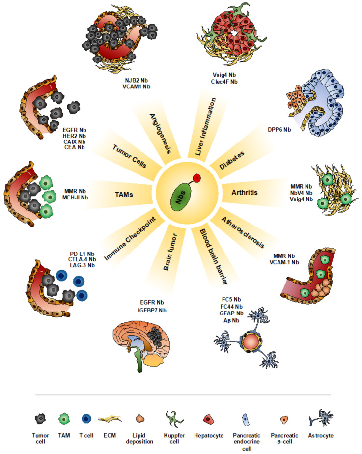
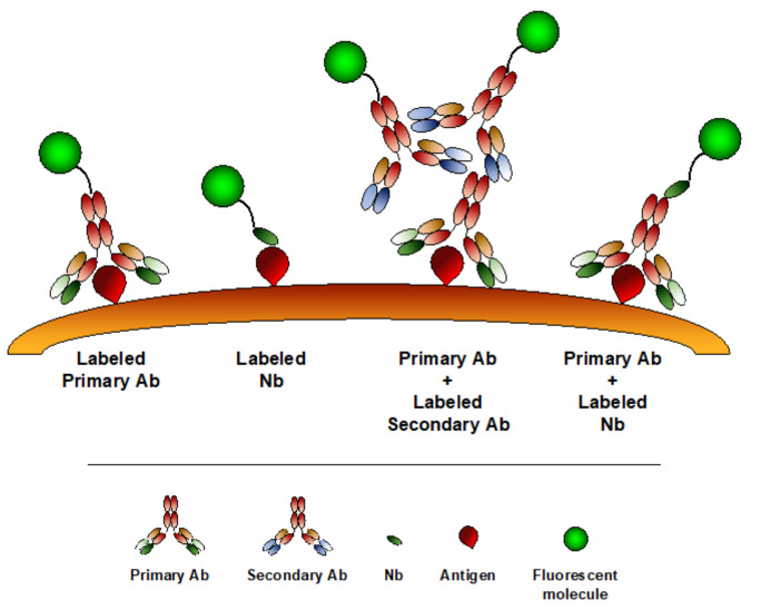
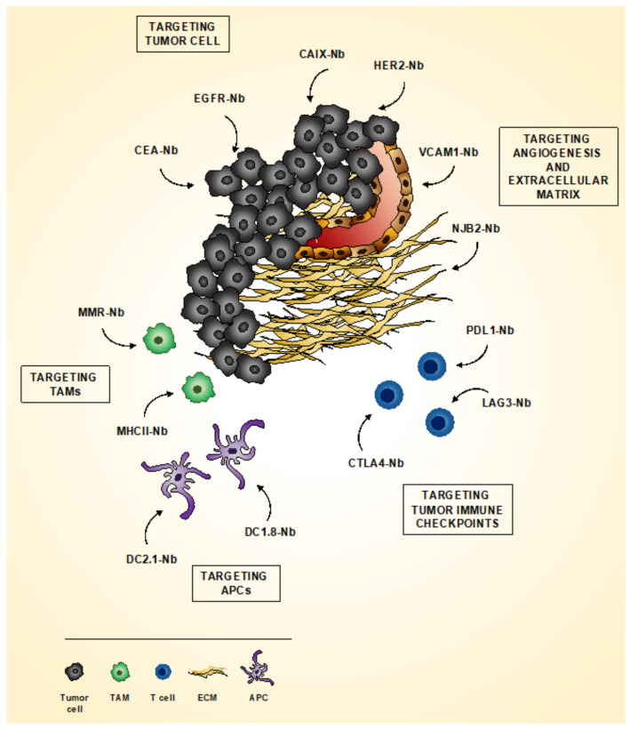
Molecular imaging is constantly growing in different areas of preclinical biomedical research. Several imaging methods have been developed and are continuously updated for both in vivo and in vitro applications, in order to increase the information about the structure, localization and function of molecules involved in physiology and disease. Along with these progresses, there is a continuous need for improving labeling strategies. In the last decades, the single domain antigen-binding fragments nanobodies (Nbs) emerged as important molecular imaging probes. Indeed, their small size (~15 kDa), high stability, affinity and modularity represent desirable features for imaging applications, providing higher tissue penetration, rapid targeting, increased spatial resolution and fast clearance. Accordingly, several Nb-based probes have been generated and applied to a variety of imaging modalities, ranging from in vivo and in vitro preclinical imaging to super-resolution microscopy. In this review, we will provide an overview of the state-of-the-art regarding the use of Nbs in several imaging modalities, underlining their extreme versatility and their enormous potential in targeting molecules and cells of interest in both preclinical and clinical studies.
分子影像学在基础医学研究的不同领域不断发展。为了增加涉及生理和疾病的分子的结构、定位和功能的信息,已经开发并不断更新了几种用于体内和体外应用的成像方法。随着这些进展,不断需要改进标记策略。在过去的几十年中,单域抗原结合片段纳米抗体(Nbs)作为重要的分子成像探针出现。事实上,它们的小尺寸(~15 kDa)、高稳定性、亲和力和模块化是成像应用的理想特征,提供了更高的组织穿透性、快速靶向、更高的空间分辨率和快速清除。因此,已经生成了几种基于 Nb 的探针,并应用于各种成像方式,从体内和体外临床前成像到超分辨率显微镜。在这篇综述中,我们将概述 Nb 在几种成像方式中的最新应用,强调它们的极端多功能性及其在靶向临床前和临床研究中感兴趣的分子和细胞方面的巨大潜力。