Department of Surgery, University of California San Diego, CA.
Laboratory for In vivo Cellular and Molecular Imaging, ICMI-BEFY/MIMA, Vrije Universiteit Brussel, Brussels, Belgium.
Surgery. 2020 Jul;168(1):85-91. doi: 10.1016/j.surg.2020.02.020. Epub 2020 May 4.
Nanobodies, derived from camelid antibodies made of only heavy chains, are the smallest, biologic, antigen-binding fragments (~15kDa) with faster pharmacokinetics and better tumor penetration efficiency than standard antibodies. The present study evaluates the efficacy of a fluorescent, anti-carcinoembryonic antigen (CEA) nanobody for rapid tumor labeling in an orthotopic mouse model of pancreatic cancer.
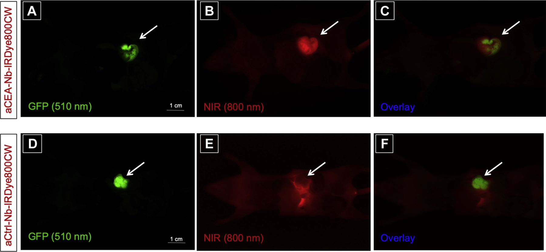
Anti-CEA or control nanobodies were conjugated with the near-infrared fluorophore IRDye 800CW. Fragments of BxPC-3 (high-CEA expressing) or MiaPACA-2 (low-CEA expressing) human pancreatic cancer cell lines were orthotopically implanted into the pancreatic tail of nude mice. After tumors reached 7 to 10 mm in size, 2 nmol anti-CEA or control nanobody-IRDye800CW were injected intravenously. Mice were imaged at various time points hours post-injection.
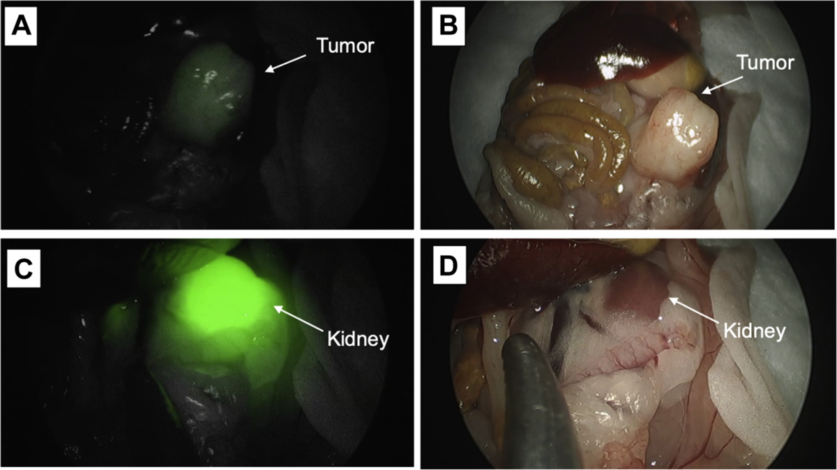
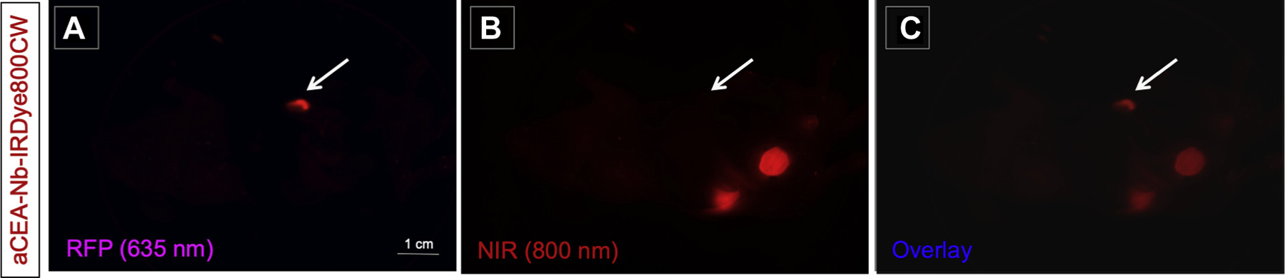
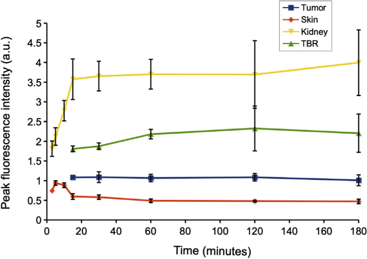
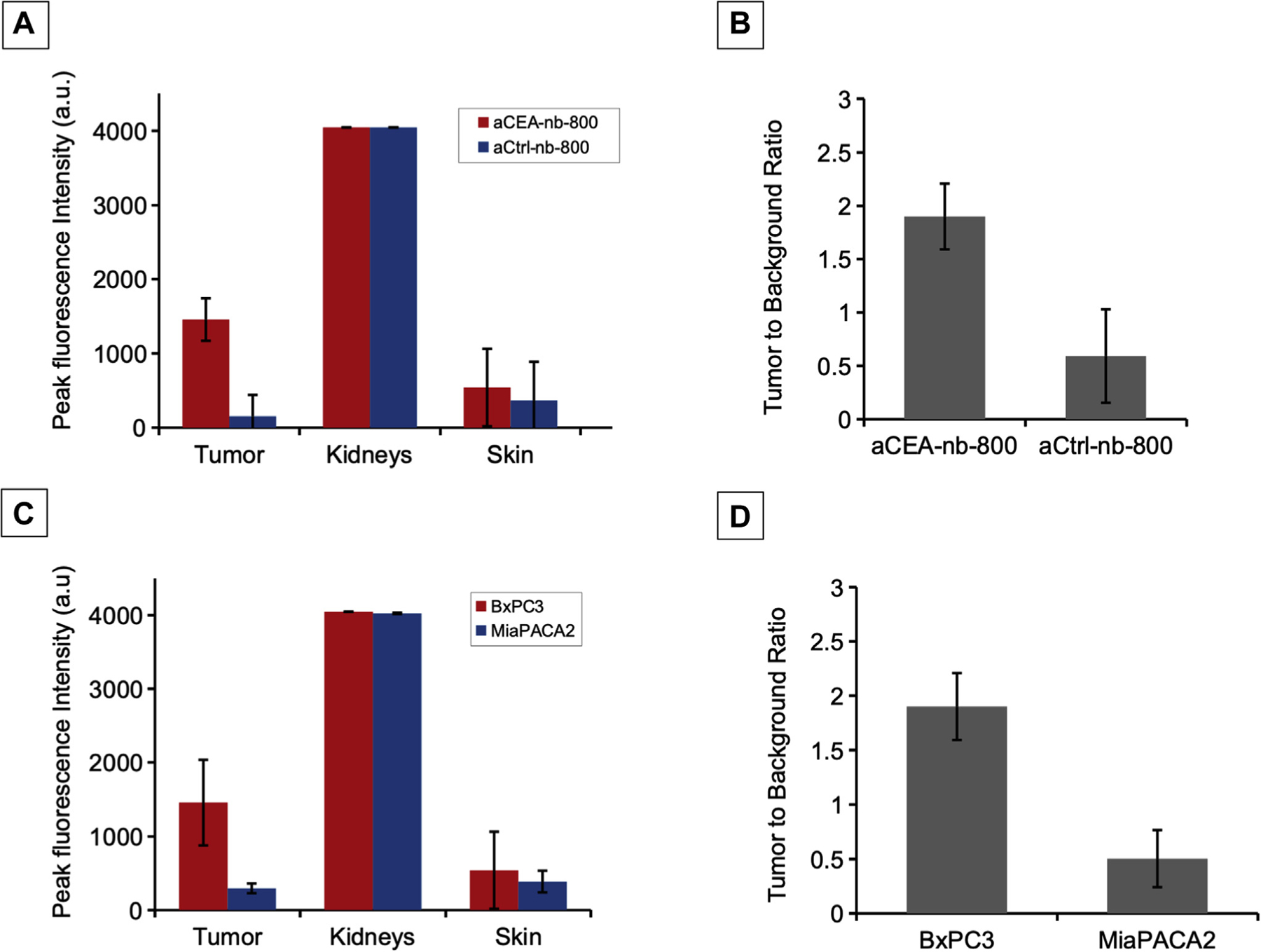
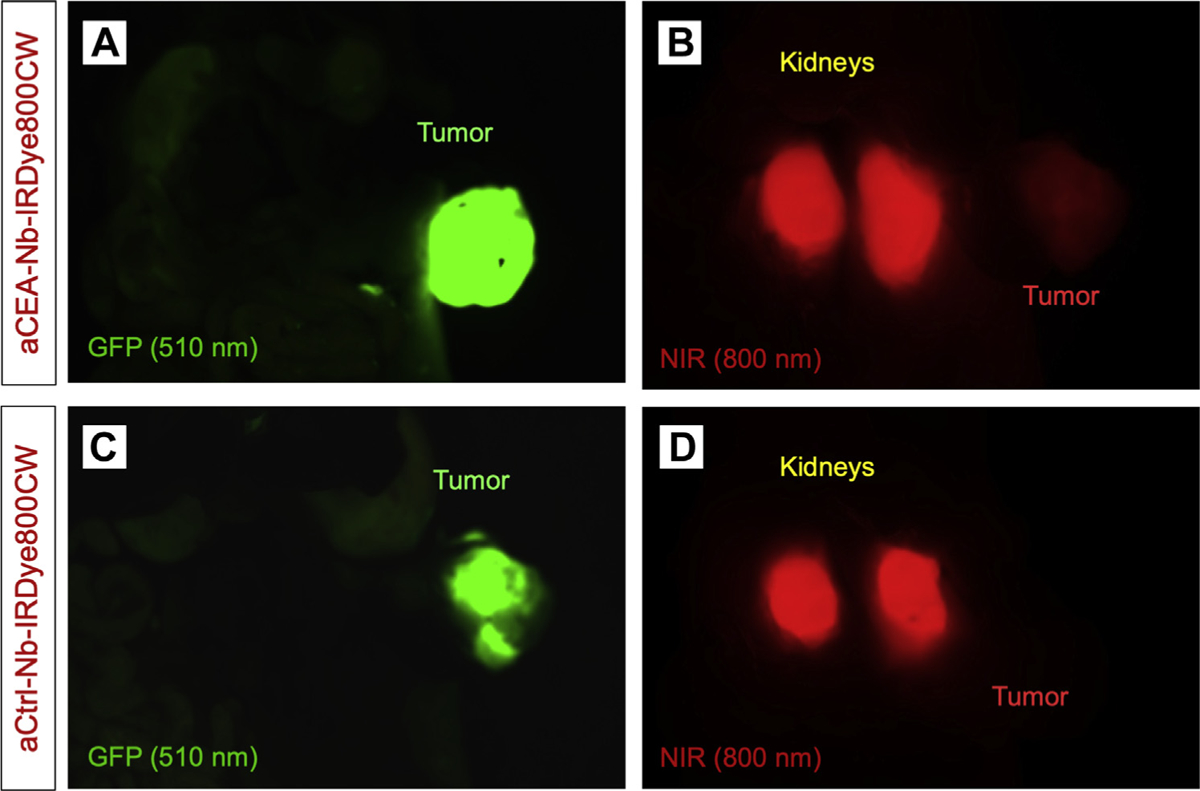
Anti-CEA nanobodies clearly labeled BxPC3 orthotopic pancreatic tumors 3 hours after injection. The signal was present as early as 15 minutes after injection and was robust at 1 to 3 hours after injection with a tumor-to-background ratio of 2.66. In contrast, there was very low accumulation in the low CEA-expressing, MiaPACA2 pancreatic orthotopic tumors. The fluorophore-conjugated nanobody was specific for CEA-expressing tumors, while the control nanobody did not show any tumor-specific signal. Both nanobodies had strong kidney uptake as expected for small-molecule probes. The fluorescence signal was detectable using 2 clinical, Food and Drug Administration-approved, 800 nm imaging devices as well as small animal imaging systems.
This anti-CEA, nanobody-based, fluorescent probe labeled pancreatic orthotopic tumors within 15 minutes of intravenous injection. Fluorescent anti-CEA nanobodies have labeling kinetics that approach the speed of nonspecific dyes such as indocyanine green but with the specificity of antibodies. The use of fluorescently-labeled, intact antibodies leads to a labeling delay of 48 to 96 hours between probe administration and the necessarily delayed time of operation, which can be avoided with nanobodies. The kinetics of a nanobody-based probe makes it a practical agent for same-day, patient administration and fluorescence-guided surgery.
纳米抗体来源于仅由重链组成的骆驼科抗体,是最小的生物抗原结合片段(~15kDa),与标准抗体相比,具有更快的药代动力学和更好的肿瘤穿透效率。本研究评估了荧光抗癌胚抗原(CEA)纳米抗体在胰腺癌细胞原位模型中快速标记肿瘤的疗效。
将抗 CEA 或对照纳米抗体与近红外荧光染料 IRDye 800CW 缀合。将 BxPC-3(高 CEA 表达)或 MiaPACA-2(低 CEA 表达)人胰腺癌细胞系的片段原位植入裸鼠胰腺尾部。当肿瘤大小达到 7 至 10mm 时,静脉内注射 2nmol 抗 CEA 或对照纳米抗体-IRDye800CW。注射后不同时间点进行小鼠成像。
抗 CEA 纳米抗体在注射后 3 小时清楚地标记了 BxPC3 原位胰腺肿瘤。在注射后 15 分钟即可检测到信号,在注射后 1 至 3 小时内信号很强,肿瘤与背景的比值为 2.66。相比之下,在低 CEA 表达的 MiaPACA2 胰腺原位肿瘤中,积累非常低。荧光染料缀合的纳米抗体是 CEA 表达肿瘤的特异性探针,而对照纳米抗体则没有显示任何肿瘤特异性信号。两种纳米抗体都对小分子探针具有强烈的肾脏摄取作用。荧光信号可使用 2 种临床批准的、美国食品和药物管理局批准的 800nm 成像设备以及小动物成像系统检测到。
这种基于抗 CEA 纳米抗体的荧光探针在静脉注射后 15 分钟内标记了胰腺原位肿瘤。荧光抗 CEA 纳米抗体的标记动力学接近非特异性染料(如吲哚菁绿)的速度,但具有抗体的特异性。使用荧光标记的完整抗体会导致探针给药与必要的手术延迟之间出现 48 至 96 小时的延迟,而这可以通过纳米抗体避免。纳米抗体探针的动力学使其成为一种实用的试剂,可用于当天患者给药和荧光引导手术。