• 文献检索
  • 文档翻译
  • 深度研究
  • 学术资讯
  • Suppr Zotero 插件Zotero 插件
  • 邀请有礼
  • 套餐&价格
  • 历史记录
应用&插件
Suppr Zotero 插件Zotero 插件浏览器插件Mac 客户端Windows 客户端微信小程序
定价
高级版会员购买积分包购买API积分包
服务
文献检索文档翻译深度研究API 文档MCP 服务
关于我们
关于 Suppr公司介绍联系我们用户协议隐私条款
关注我们

Suppr 超能文献

核心技术专利:CN118964589B侵权必究
粤ICP备2023148730 号-1Suppr @ 2026

文献检索

告别复杂PubMed语法,用中文像聊天一样搜索,搜遍4000万医学文献。AI智能推荐,让科研检索更轻松。

立即免费搜索

文件翻译

保留排版,准确专业,支持PDF/Word/PPT等文件格式,支持 12+语言互译。

免费翻译文档

深度研究

AI帮你快速写综述,25分钟生成高质量综述,智能提取关键信息,辅助科研写作。

立即免费体验

应用超声造影监测肝细胞癌大鼠模型中靶向治疗的疗效:重点探讨扫描仪间和操作者间的可重复性。

Therapeutic response monitoring after targeted therapy in an orthotopic rat model of hepatocellular carcinoma using contrast-enhanced ultrasound: Focusing on inter-scanner, and inter-operator reproducibility.

机构信息

Department of Radiology, Pusan National University Yangsan Hospital, Yangsan-si, Gyeongsangnam-do, Korea.

Department of Radiology, Seoul National University Hospital, Jongno-gu, Seoul, Korea.

出版信息

PLoS One. 2020 Dec 23;15(12):e0244304. doi: 10.1371/journal.pone.0244304. eCollection 2020.

DOI:10.1371/journal.pone.0244304
PMID:33362203
原文链接:https://pmc.ncbi.nlm.nih.gov/articles/PMC7757904/
Abstract

PURPOSE

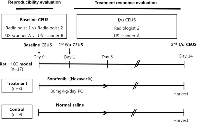
To assess therapeutic response monitoring after targeted therapy in an orthotopic rat model of hepatocellular carcinoma (HCC) using CEUS with focusing on inter-scanner and inter-operator reproducibility.

MATERIALS AND METHODS

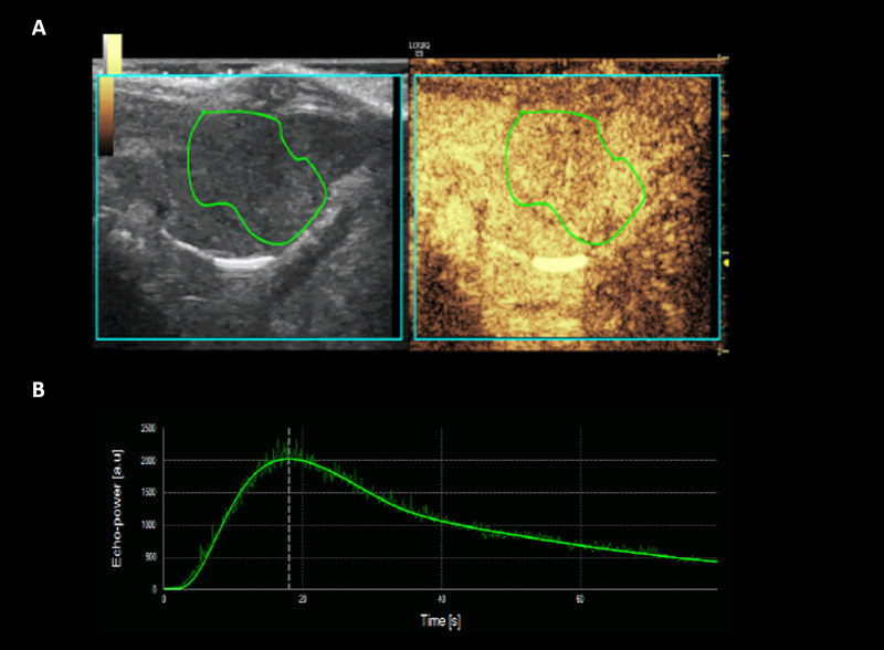
For reproducibility, CEUS was performed using two different US scanners by two operators in sixteen rat models of HCC. Using perfusion analysis software (VueBox ®), eleven parameters were collected, and intra-class correlation coefficient (ICC) was used to analyze reproducibility. Then seventeen rat models of HCC were divided into treatment group (n = 8, 30 mg/kg/day sorafenib for five days) and control group (n = 9). CEUS was performed at baseline and 14 days after first treatment, and changes of perfusion parameters were analyzed.

RESULTS

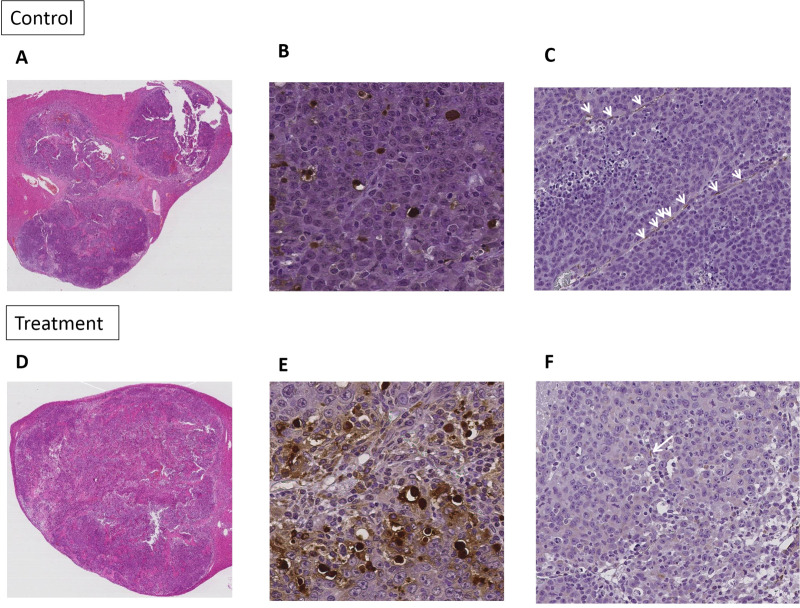
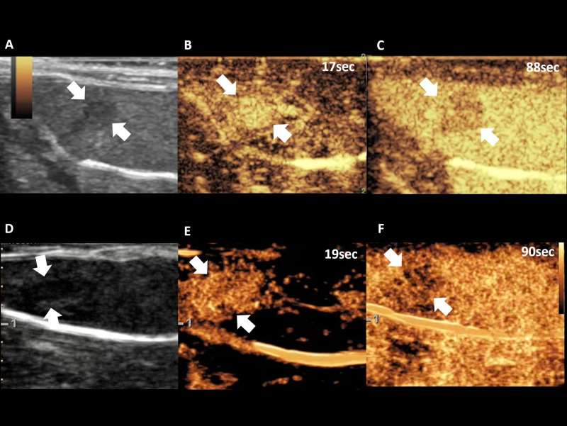
In treatment group, CEUS perfusion parameters showed a significant change. The peak enhancement (PE, 2.50 x10³±1.68 x10³ vs 5.55x10²±4.65x10², p = 0.010) and wash-in and wash out AUC (WiWoAUC, 1.07x10⁵±6.48 x10⁴ vs 2.65x10⁴±2.25x10⁴, p = 0.009) had significantly decreased two weeks after treatment. On the contrary, control group did not show a significant change, including PE (1.15 x10³±7.53x10² vs 9.43x10²± 7.81 x10², p = 0.632) and WiWoAUC (5.09 x10⁴±3.25x10⁴ vs 5.92 x10⁴±3.20x10⁴, p = 0.646). For reproducibility, the various degrees of inter-scanner reproducibility were from poor to good (ICC: <0.01-0.63). However, inter-operator reproducibility of important perfusion parameters, including WiAUC, WoAUC, and WiWoAUC, ranged from fair to excellent (ICC: 0.59-0.93) in a different scanner.

CONCLUSION

Our results suggest that CEUS is useful for assessment of the treatment response after targeted therapy and with fair to excellent inter-operator reproducibility.

摘要

目的

使用超声造影(CEUS)评估两种不同超声扫描仪和两名操作者在肝癌(HCC)大鼠模型中的靶向治疗后疗效监测的可重复性。

材料与方法

为了评估可重复性,16 只 HCC 大鼠模型分别由两名操作者使用两台不同的超声扫描仪进行 CEUS。使用灌注分析软件(VueBox ®)收集 11 个参数,采用组内相关系数(ICC)分析其可重复性。然后,将 17 只 HCC 大鼠模型分为治疗组(n=8,每天 30mg/kg 索拉非尼治疗 5 天)和对照组(n=9)。在基线和首次治疗后 14 天进行 CEUS,并分析灌注参数的变化。

结果

在治疗组中,CEUS 灌注参数显示出显著变化。治疗后两周,峰值增强(PE,2.50x103±1.68x10³ 比 5.55x10²±4.65x10²,p=0.010)和流入和流出面积(WiWoAUC,1.07x105±6.48x10⁴ 比 2.65x10⁴±2.25x10⁴,p=0.009)显著降低。相反,对照组没有显示出明显的变化,包括 PE(1.15x10³±7.53x10² 比 9.43x10²±7.81x10²,p=0.632)和 WiWoAUC(5.09x10⁴±3.25x10⁴ 比 5.92x10⁴±3.20x10⁴,p=0.646)。对于可重复性,不同程度的扫描仪之间的可重复性从差到好(ICC:<0.01-0.63)。然而,在不同的扫描仪中,WiAUC、WoAUC 和 WiWoAUC 等重要灌注参数的操作者之间的可重复性从一般到优秀(ICC:0.59-0.93)。

结论

我们的研究结果表明,CEUS 可用于评估靶向治疗后的治疗反应,且具有良好到优秀的操作者间可重复性。

https://cdn.ncbi.nlm.nih.gov/pmc/blobs/0a47/7757904/5da55f523b99/pone.0244304.g004.jpg
https://cdn.ncbi.nlm.nih.gov/pmc/blobs/0a47/7757904/e19a104ab82c/pone.0244304.g001.jpg
https://cdn.ncbi.nlm.nih.gov/pmc/blobs/0a47/7757904/8f674e571987/pone.0244304.g002.jpg
https://cdn.ncbi.nlm.nih.gov/pmc/blobs/0a47/7757904/425848e7e87b/pone.0244304.g003.jpg
https://cdn.ncbi.nlm.nih.gov/pmc/blobs/0a47/7757904/5da55f523b99/pone.0244304.g004.jpg

相似文献

1
Therapeutic response monitoring after targeted therapy in an orthotopic rat model of hepatocellular carcinoma using contrast-enhanced ultrasound: Focusing on inter-scanner, and inter-operator reproducibility.应用超声造影监测肝细胞癌大鼠模型中靶向治疗的疗效:重点探讨扫描仪间和操作者间的可重复性。
PLoS One. 2020 Dec 23;15(12):e0244304. doi: 10.1371/journal.pone.0244304. eCollection 2020.
2
Potential application of dynamic contrast enhanced ultrasound in predicting microvascular invasion of hepatocellular carcinoma.动态对比增强超声在预测肝细胞癌微血管侵犯中的应用潜力。
Clin Hemorheol Microcirc. 2021;77(4):461-469. doi: 10.3233/CH-201085.
3
Savitzky-Golay filter based contrast-enhanced ultrasound quantification in hepatic tumors: Methodology and its correlation with tumor angiogenesis.基于 Savitzky-Golay 滤波器的肝肿瘤超声增强定量法:方法学及其与肿瘤血管生成的相关性。
Clin Hemorheol Microcirc. 2019;73(2):271-282. doi: 10.3233/CH-180432.
4
Comparison of dynamic contrast-enhanced magnetic resonance imaging and contrast-enhanced ultrasound for evaluation of the effects of sorafenib in a rat model of hepatocellular carcinoma.动态对比增强磁共振成像与超声造影在评价索拉非尼对大鼠肝细胞癌模型疗效中的比较
Magn Reson Imaging. 2019 Apr;57:156-164. doi: 10.1016/j.mri.2018.11.012. Epub 2018 Nov 19.
5
Dynamic contrast-enhanced ultrasound (DCE-US) for the characterization of hepatocellular carcinoma and cholangiocellular carcinoma.动态对比增强超声(DCE-US)用于肝细胞癌和胆管细胞癌的特征描述。
Ultraschall Med. 2014 Dec;35(6):522-7. doi: 10.1055/s-0034-1385170. Epub 2014 Sep 9.
6
The value of contrast-enhanced ultrasound quantitative parameters in the prognosis prediction of hepatocellular carcinoma after thermal ablation: a retrospective cohort study.超声造影定量参数在热消融术后肝细胞癌预后预测中的价值:一项回顾性队列研究
J Gastrointest Oncol. 2022 Oct;13(5):2522-2531. doi: 10.21037/jgo-22-919.
7
Parametric imaging with contrast-enhanced ultrasound for differentiating hepatocellular carcinoma from metastatic liver cancer.超声造影参数成像在鉴别肝细胞癌与肝转移癌中的应用
Clin Hemorheol Microcirc. 2016 Nov 25;64(2):177-188. doi: 10.3233/CH-162060.
8
Quantitative perfusion analysis of hepatocellular carcinoma using dynamic contrast enhanced ultrasound (CEUS) to determine tumor microvascularization.使用动态对比增强超声(CEUS)定量分析肝细胞癌以确定肿瘤微血管化。
Clin Hemorheol Microcirc. 2019;73(1):95-104. doi: 10.3233/CH-199221.
9
Renal elasticity and perfusion changes on ultrasonography in an early-stage diabetic rat model.早期糖尿病大鼠模型超声检查中肾脏弹性和灌注的变化
Quant Imaging Med Surg. 2023 Dec 1;13(12):7667-7679. doi: 10.21037/qims-23-409. Epub 2023 Oct 13.
10
Color Coded Perfusion Imaging with Contrast Enhanced Ultrasound (CEUS) for Post-Interventional Success Control Following Irreversible Electroporation (IRE) of Primary and Secondary Malignant Liver Lesions.对比增强超声(CEUS)彩色编码灌注成像用于原发性和继发性恶性肝脏病变不可逆电穿孔(IRE)后的介入后成功控制。
J Gastrointestin Liver Dis. 2019 Sep 1;28(3):311-318. doi: 10.15403/jgld-254.

引用本文的文献

1
A Comprehensive and Repeatable Contrast-Enhanced Ultrasound Quantification Approach for Clinical Evaluations of Tumor Blood Flow.一种用于肿瘤血流临床评估的全面且可重复的超声造影定量方法。
Invest Radiol. 2025 Apr 1;60(4):281-290. doi: 10.1097/RLI.0000000000001127. Epub 2024 Oct 9.
2
Quantitative analysis of contrast-enhanced ultrasonography in rat models of hepatic acute graft-versus-host disease.肝急性移植物抗宿主病大鼠模型中超声造影的定量分析
Quant Imaging Med Surg. 2023 Aug 1;13(8):4908-4918. doi: 10.21037/qims-22-1145. Epub 2023 May 22.

本文引用的文献

1
Evaluation of the Reproducibility of Bolus Transit Quantification With Contrast-Enhanced Ultrasound Across Multiple Scanners and Analysis Software Packages-A Quantitative Imaging Biomarker Alliance Study.对比增强超声在多个扫描仪和分析软件包中对灌注定量的重复性评估——定量成像生物标志物联盟研究。
Invest Radiol. 2020 Oct;55(10):643-656. doi: 10.1097/RLI.0000000000000702.
2
Papillary renal cell carcinoma in contrast-enhanced ultrasound (CEUS) - A diagnostic performance study.超声造影(CEUS)在乳头状肾细胞癌中的应用——一项诊断性能研究。
Clin Hemorheol Microcirc. 2019;71(2):159-164. doi: 10.3233/CH-189406.
3
Contrast Enhanced Three Dimensional Ultrasound for Intraluminal Thrombus Assessment in Abdominal Aortic Aneurysms.
对比增强三维超声在腹主动脉瘤管腔内血栓评估中的应用。
Eur J Vasc Endovasc Surg. 2018 Nov;56(5):673-680. doi: 10.1016/j.ejvs.2018.07.029. Epub 2018 Aug 27.
4
Non-invasive monitoring of the therapeutic response in sorafenib-treated hepatocellular carcinoma based on photoacoustic imaging.基于光声成像的索拉非尼治疗肝细胞癌的疗效无创监测。
Eur Radiol. 2018 Jan;28(1):372-381. doi: 10.1007/s00330-017-4960-3. Epub 2017 Jul 27.
5
The value of contrast-enhanced ultrasound (CEUS) using a high-end ultrasound system in the characterization of endoleaks after endovascular aortic repair (EVAR).使用高端超声系统的对比增强超声(CEUS)在血管腔内主动脉修复术(EVAR)后内漏特征分析中的价值。
Clin Hemorheol Microcirc. 2017;66(4):283-292. doi: 10.3233/CH-179102.
6
Time-Intensity Curves Obtained after Microbubble Injection Can Be Used to Differentiate Responders from Nonresponders among Patients with Clinically Active Crohn Disease after 6 Weeks of Pharmacologic Treatment.经微泡注射后获得的时间-强度曲线可用于区分在药物治疗 6 周后患有临床活动性克罗恩病的患者中的应答者和无应答者。
Radiology. 2016 Nov;281(2):606-616. doi: 10.1148/radiol.2016152461. Epub 2016 May 18.
7
Reproducibility of Contrast-Enhanced Ultrasound in Mice with Controlled Injection.控制注射条件下小鼠对比增强超声的可重复性
Mol Imaging Biol. 2016 Oct;18(5):651-8. doi: 10.1007/s11307-016-0952-y.
8
Simultaneous Assessment of Myocardial Perfusion, Wall Motion, and Deformation during Myocardial Contrast Echocardiography: A Feasibility Study.心肌对比超声心动图中同时评估心肌灌注、壁运动和变形:一项可行性研究。
Echocardiography. 2016 Jun;33(6):889-95. doi: 10.1111/echo.13190. Epub 2016 Feb 1.
9
Early Clinical Response after 2 Weeks of Sorafenib Therapy Predicts Outcomes and Anti-Tumor Response in Patients with Advanced Hepatocellular Carcinoma.索拉非尼治疗2周后的早期临床反应可预测晚期肝细胞癌患者的预后和抗肿瘤反应。
PLoS One. 2015 Sep 30;10(9):e0138776. doi: 10.1371/journal.pone.0138776. eCollection 2015.
10
Metrology Standards for Quantitative Imaging Biomarkers.定量成像生物标志物的计量标准。
Radiology. 2015 Dec;277(3):813-25. doi: 10.1148/radiol.2015142202. Epub 2015 Aug 12.