Clearside Biomedical, Inc., Alpharetta, GA, USA.
Copernicus Therapeutics, Inc., Cleveland, OH, USA.
Transl Vis Sci Technol. 2020 Dec 15;9(13):21. doi: 10.1167/tvst.9.13.21. eCollection 2020 Dec.
This study evaluated ocular tolerability and transfectability of nonviral DNA nanoparticles (DNPs) after microneedle-based suprachoroidal (SC) administration, in comparison to subretinal (SR) administration.
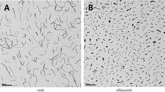
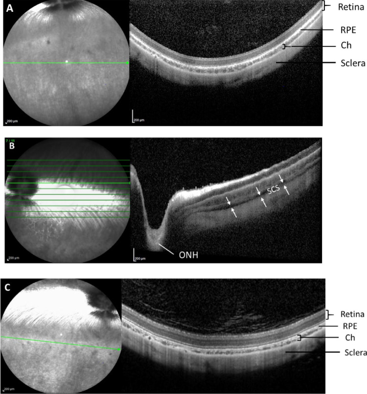
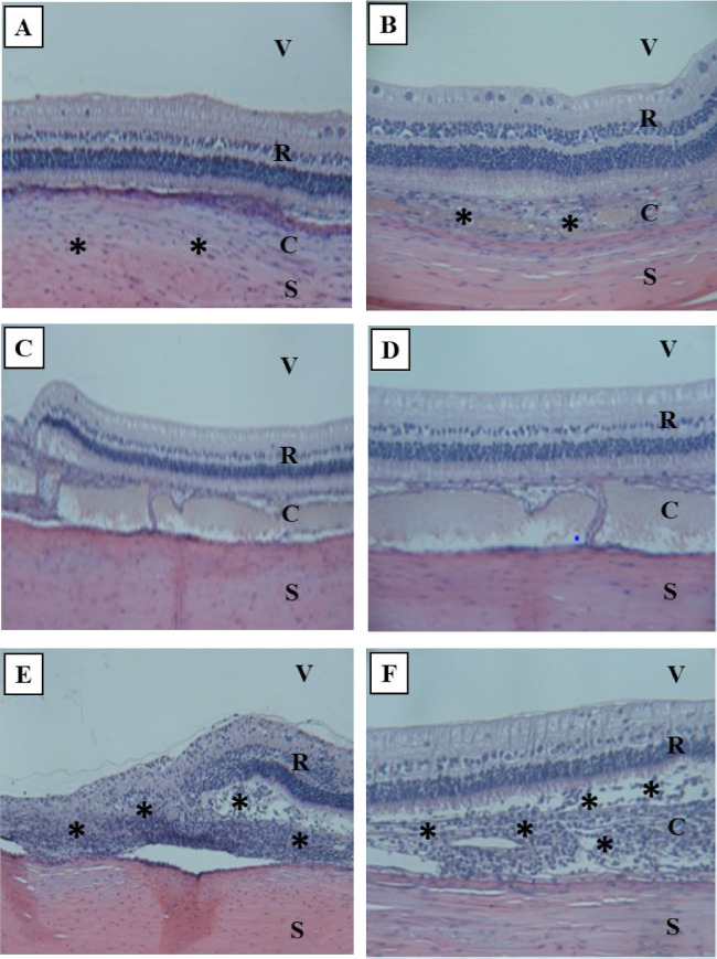
The DNPs consisted of a single copy of plasmid DNA with a polyubiquitin C/luciferase transcriptional cassette compacted with 10 kDa PEG-substituted lysine 30-mer peptides (CK30PEG10k). New Zealand White rabbits ( = 4 per group) received a unilateral SC injection (0.1 mL via a microneedle technique) of ellipsoid-shaped DNPs, rod-shaped DNPs, or saline (negative control). A cohort of rabbits ( = 4) also received a single unilateral SR injection (0.05 mL via a transvitreal approach) of rod-shaped DNPs. At day 7, luciferase activity was measured in the retina and retinal pigment epithelium (RPE)-choroid via bioluminescence assay. A cohort of rabbits received a SC injection of analogous DNPs to assess spread of DNP injectate in the suprachoroidal space (SCS) via optical coherent tomography and histology.
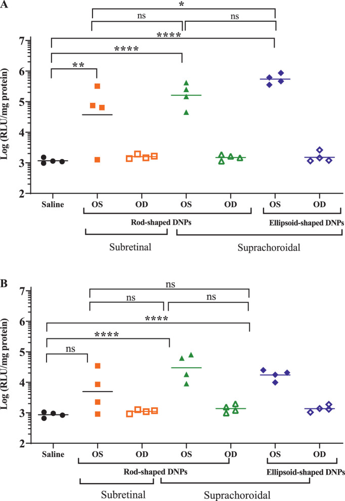
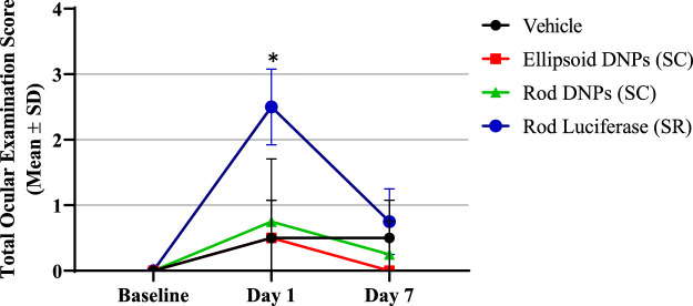
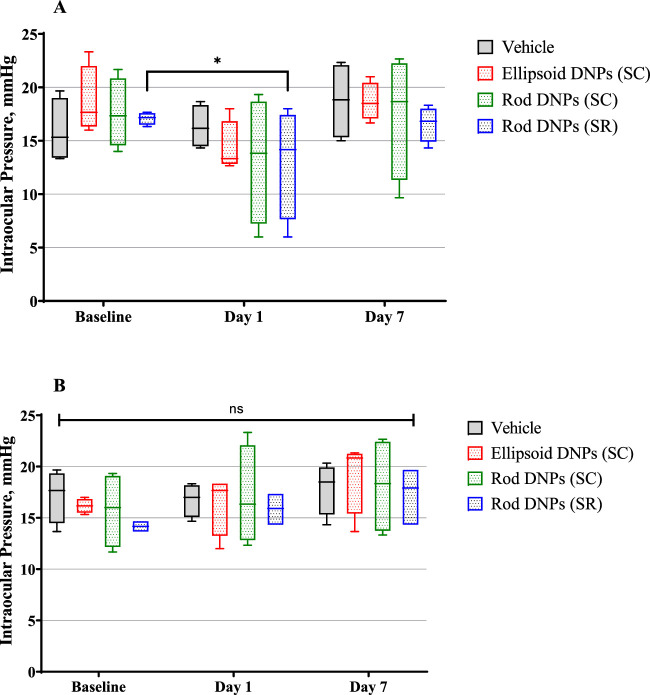
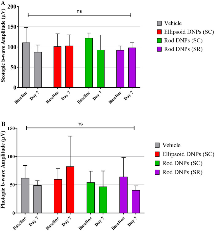
Suprachoroidal injection of DNPs resulted in reversible opening of the SCS circumferentially and posteriorly and was generally well tolerated, with no significant ocular examination score changes, intraocular pressure abnormalities, or changes in electroretinography amplitudes on day 7 compared to the baseline. High luciferase activity was observed in the retina and RPE-choroid of eyes that received SC DNPs (rod and ellipsoid shape) and SR DNPs (rod shape) compared to controls. The mean luciferase activity in RPE-choroid and retina was comparable between SC and SR administrations. Transfection in the RPE-choroid was approximately 10-fold higher than in the retina after either SC or SR administration of DNPs.
Suprachoroidal and SR administration of DNPs resulted in comparable transfection of retina and RPE-choroid.
Suprachoroidal delivery of DNPs offers the potential to precisely target chorioretinal tissues while avoiding surgical risks associated with SR injection, and it may offer an office-based nonsurgical gene therapy option for the treatment of retinal diseases.
本研究通过对比分析,评估了基于微针的脉络膜下(SC)给药与视网膜下(SR)给药后,非病毒 DNA 纳米颗粒(DNPs)的眼部耐受性和转染能力。
DNPs 由单拷贝的质粒 DNA 与多聚泛素 C/荧光素酶转录盒组成,并用 10 kDa 的聚乙二醇取代的赖氨酸 30 肽(CK30PEG10k)进行压缩。新西兰白兔(每组 4 只)接受单侧 SC 注射(通过微针技术注射 0.1 mL),分别给予类椭圆 DNPs、类杆状 DNPs 或生理盐水(阴性对照)。一组兔子(4 只)还接受了单侧 SR 注射(经玻璃体腔注射 0.05 mL)的杆状 DNPs。第 7 天,通过生物发光测定法测量视网膜和视网膜色素上皮(RPE)-脉络膜中的荧光素酶活性。一组兔子接受了类似的 DNPs 的 SC 注射,以通过光学相干断层扫描和组织学评估 DNPs 注射剂在脉络膜上腔(SCS)中的扩散情况。
SC 注射 DNPs 可使 SCS 周向和后部可逆性开放,通常耐受性良好,与基线相比,第 7 天无明显的眼部检查评分变化、眼内压异常或视网膜电图振幅变化。与对照组相比,接受 SC DNPs(类杆状和类椭圆状)和 SR DNPs(类杆状)的眼内,视网膜和 RPE-脉络膜中均观察到高荧光素酶活性。SC 和 SR 给药后,RPE-脉络膜和视网膜中的平均荧光素酶活性相似。无论是 SC 还是 SR 给药后,RPE-脉络膜中的转染效率均比视网膜高约 10 倍。
SC 和 SR 给药后,视网膜和 RPE-脉络膜的转染效率相当。
本译文仅供参考,如需使用,请以原文为准。