Kairemo Kalevi, Kappadath S Cheenu, Joensuu Timo, Macapinlac Homer A
Department of Theragnostics, Docrates Cancer Center, 00180 Helsinki, Finland.
Department of Nuclear Medicine, MD Anderson Cancer Center, Houston, TX 77030, USA.
Diagnostics (Basel). 2020 Dec 24;11(1):17. doi: 10.3390/diagnostics11010017.
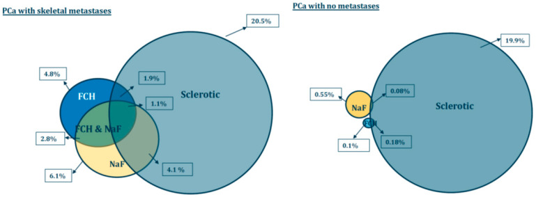
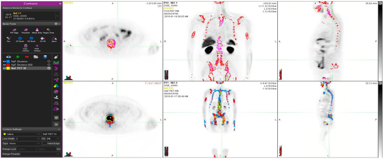
Bone metastases are common in prostate cancer (PCa). Fluorocholine-18 (FCH) and sodium fluoride-18 (NaF) have been used to assess PCa associated skeletal disease in thousands of patients by demonstrating different mechanism of uptake-cell membrane (lipid) synthesis and bone mineralization. Here, this difference is characterized quantitatively in detail. Our study cohort consisted of 12 patients with advanced disease (> 5 lesions) (M) and of five PCa patients with no skeletal disease (N). They had routine PET/CT with FCH and NaF on consecutive days. Skeletal regions in CT were used to co-register the two PET/CT scans. Bone 3-D volume of interest (VOI) was defined on the CT of PET with a threshold of HU > 150, and sclerotic/dense bone as HU > 600, respectively. Additional VOIs were defined on PET uptake with the threshold values on both FCH (SUV > 3.5) and NaF (SUV > 10). The pathologic skeletal volumes for each technique (CT, HU > 600), NaF (SUV > 10) and FCH (SUV > 3.5) were developed and analyzed. The skeletal VOIs varied from 5.03 L to 7.31 L, whereas sclerotic bone VOIs were from 0.88 L to 2.99 L. Total choline kinase (cell membrane synthesis) activity for FCH (TCA) varied from 0.008 to 4.85 [kg] in M group and from 0.0006 to 0.085 [kg] in N group. Total accelerated osteoblastic (bone demineralization) activity for NaF (TBA varied from 0.25 to 13.6 [kg] in M group and varied from 0.000 to 1.09 [kg] in N group. The sclerotic bone volume represented only 1.86 ± 1.71% of the pathologic FCH volume and 4.07 ± 3.21% of the pathologic NaF volume in M group, and only 0.08 ± 0.09% and 0.18 ± 0.19% in N group, respectively. Our results suggest that CT alone cannot be used for the assessment of the extent of active metastatic skeletal disease in PCa. NaF and FCH give complementary information about the activity of the skeletal disease, improving diagnosis and disease staging.
骨转移在前列腺癌(PCa)中很常见。氟胆碱 - 18(FCH)和氟化钠 - 18(NaF)已被用于通过展示不同的摄取机制——细胞膜(脂质)合成和骨矿化,来评估数千例与PCa相关的骨骼疾病。在此,详细定量地描述了这种差异。我们的研究队列包括12例晚期疾病(> 5个病灶)患者(M组)和5例无骨骼疾病的PCa患者(N组)。他们在连续两天进行了FCH和NaF的常规PET/CT检查。CT中的骨骼区域用于对两次PET/CT扫描进行配准。在PET的CT上,以HU > 150为阈值定义骨三维感兴趣区(VOI),分别以HU > 600定义硬化/致密骨。在FCH(SUV > 3.5)和NaF(SUV > 10)的阈值下,在PET摄取上定义额外的VOI。开发并分析了每种技术(CT,HU > 600)、NaF(SUV > 10)和FCH(SUV > 3.5)的病理性骨骼体积。骨骼VOI从5.03 L到7.31 L不等,而硬化骨VOI从0.88 L到2.99 L不等。M组中FCH的总胆碱激酶(细胞膜合成)活性(TCA)从0.008到4.85 [kg]不等,N组从0.0006到0.085 [kg]不等。M组中NaF的总加速成骨细胞(骨脱矿质)活性(TBA)从0.25到13.6 [kg]不等,N组从0.000到1.09 [kg]不等。在M组中,硬化骨体积仅占病理性FCH体积的1.86 ± 1.71%和病理性NaF体积的4.07 ± 3.21%,在N组中分别仅占0.08 ± 0.09%和0.18 ± 0.19%。我们的结果表明,仅CT不能用于评估PCa中活跃转移性骨骼疾病的范围。NaF和FCH提供了关于骨骼疾病活动的互补信息,改善了诊断和疾病分期。