Lee Ju-Young, Choung Han-Wool, Choung Pill-Hoon
Department of Oral and Maxillofacial Surgery, Seoul St. Mary's Hospital, Seoul, Korea.
Department of Oral and Maxillofacial Surgery, Dental Research Institute, School of Dentistry, Seoul National University, Seoul, Korea.
J Korean Assoc Oral Maxillofac Surg. 2020 Dec 31;46(6):379-384. doi: 10.5125/jkaoms.2020.46.6.379.
We sought to identify a clinically useful method of analyzing orbital dystopia to aid in diagnosis and treatment planning and to quantify vertical discrepancies in eye level and variations in canthal tilt in Koreans.
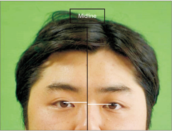
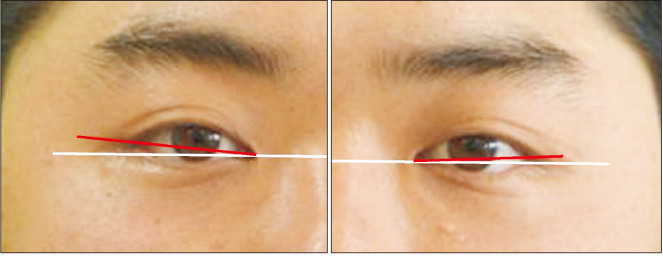
In 76 Korean patients with a mean age of 23.12 years, mean differences in the level of the pupils, lateral canthi, medial canthi, and canthal tilt were measured. The difference in pupil level was calculated from the perpendicular lines drawn from the midpupil area of each eye to the midline of the face to determine the amount of skeletal discrepancy of the eye. Soft tissue discrepancies were determined according to the vertical difference between the lines drawn from the lateral or medial canthus of each eye perpendicular to the midline of the face. The canthal tilt was determined from the inclination of a line connecting the lateral and medial canthi, then classified as class I, II, or III.
Mean differences in pupil level, medial canthi, and lateral canthi were 1.57±1.10 mm, 1.14±1.07 mm, and 2.03±1.64 mm, respectively. The mean degree of canthal tilt were 8.45°±3.53° for the right side and 8.42°±3.81° for the left side. No study participants presented with class III canthal tilt. The mean canthal tilt values for those with class I tilt were 3.21°±1.68° for the right side and 3.18°±1.63° for the left side, while, for those who had class II tilt, the values were 9.60°±3.66° for the right side and 9.54°±2.99° for the left side.
The presented diagnostic method of orbital dystopia can be used to effectively establish a treatment plan that takes into consideration the patient's skeletal and soft-tissue discrepancies.
我们试图确定一种临床上有用的分析眼眶错位的方法,以辅助诊断和治疗规划,并量化韩国人眼位的垂直差异和眦倾斜度的变化。
对76例平均年龄为23.12岁的韩国患者,测量瞳孔水平、外眦、内眦和眦倾斜度的平均差异。通过从每只眼睛瞳孔中点区域向面部中线绘制垂直线来计算瞳孔水平差异,以确定眼睛的骨骼差异量。软组织差异根据从每只眼睛的外眦或内眦向面部中线绘制的线之间的垂直差异来确定。眦倾斜度由连接外眦和内眦的线的倾斜度确定,然后分为I类、II类或III类。
瞳孔水平、内眦和外眦的平均差异分别为1.57±1.10mm、1.14±1.07mm和2.03±1.64mm。右侧眦倾斜度的平均度数为8.45°±3.53°,左侧为8.42°±3.81°。没有研究参与者表现为III类眦倾斜。I类倾斜者右侧眦倾斜度的平均值为3.21°±1.68°,左侧为3.18°±1.63°;而II类倾斜者右侧的值为9.60°±3.66°,左侧为9.54°±2.99°。
所提出的眼眶错位诊断方法可用于有效制定考虑患者骨骼和软组织差异的治疗计划。