• 文献检索
  • 文档翻译
  • 深度研究
  • 学术资讯
  • Suppr Zotero 插件Zotero 插件
  • 邀请有礼
  • 套餐&价格
  • 历史记录
应用&插件
Suppr Zotero 插件Zotero 插件浏览器插件Mac 客户端Windows 客户端微信小程序
定价
高级版会员购买积分包购买API积分包
服务
文献检索文档翻译深度研究API 文档MCP 服务
关于我们
关于 Suppr公司介绍联系我们用户协议隐私条款
关注我们

Suppr 超能文献

核心技术专利:CN118964589B侵权必究
粤ICP备2023148730 号-1Suppr @ 2026

文献检索

告别复杂PubMed语法,用中文像聊天一样搜索,搜遍4000万医学文献。AI智能推荐,让科研检索更轻松。

立即免费搜索

文件翻译

保留排版,准确专业,支持PDF/Word/PPT等文件格式,支持 12+语言互译。

免费翻译文档

深度研究

AI帮你快速写综述,25分钟生成高质量综述,智能提取关键信息,辅助科研写作。

立即免费体验

既往活检是否会导致浅表非壶腹十二指肠上皮肿瘤的癌症过度诊断?

Does previous biopsy lead to cancer overdiagnosis of superficial non-ampullary duodenal epithelial tumors?

作者信息

Tsuji Shigetsugu, Doyama Hisashi, Tsuyama Sho, Dejima Akihiro, Nakashima Takashi, Wakita Shigenori, Kito Yosuke, Nakanishi Hiroyoshi, Yoshida Naohiro, Katayanagi Kazuyoshi, Minato Hiroshi, Yao Takashi, Yao Kenshi

机构信息

Department of Gastroenterology, Ishikawa Prefectural Central Hospital, Kanazawa, Japan.

Department of Human Pathology, Graduate School of Medicine, Juntendo University, Tokyo, Japan.

出版信息

Endosc Int Open. 2021 Jan;9(1):E58-E65. doi: 10.1055/a-1293-7487. Epub 2021 Jan 1.

DOI:10.1055/a-1293-7487
PMID:33403237
原文链接:https://pmc.ncbi.nlm.nih.gov/articles/PMC7775803/
Abstract

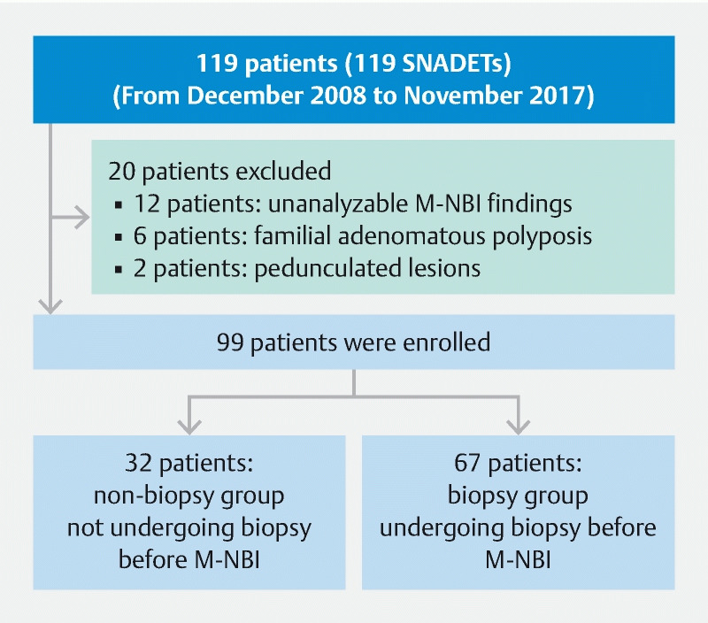
We aimed to evaluate the diagnostic performance of magnifying endoscopy with narrow-band imaging (M-NBI) in superficial non-ampullary duodenal epithelial tumors (SNADETs) regarding the absence or presence of biopsy before M-NBI diagnosis.  Clinicopathological data were retrospectively reviewed for 99 SNADETs from 99 patients who underwent endoscopic resection. The 99 tumors were divided into the non-biopsy group (32 lesions not undergoing biopsy before M-NBI examination) and the biopsy group (67 lesions undergoing biopsy before M-NBI examination). We investigated the correlation between the M-NBI diagnosis and the histopathological diagnosis of the SNADETs in both groups.  According to the modified revised Vienna classification, 31 tumors were classified as category 3 (C3) (low-grade adenoma) and 68 as category 4/5 (C4/5) (high-grade adenoma/cancer). The accuracy, sensitivity, and specificity of preoperative M-NBI diagnoses in the non-biopsy group vs the biopsy group were 88 % (95 % confidence interval: 71.0 - 96.5) vs 66 % (51.5 - 75.5),  = 0.02; 95 % (77.2 - 99.9) vs 89 % (76.4 - 96.4),  = 0.39; and 70 % (34.8 - 93.3) vs 14 % (3.0 - 36.3),  < 0.01, respectively. Notably, in the biopsy group, the specificity of M-NBI in SNADETs was low at only 14 % because we over-diagnosed most C3 lesions as C4/5. M-NBI findings might have been compromised by the previous biopsy procedure itself.  In the non-biopsy group, the accuracy of M-NBI in SNADETs was excellent in distinguishing C4/5 lesions from C3. The M-NBI findings in SNADETs should be evaluated while carefully considering the influence of a previous biopsy.

摘要

我们旨在评估窄带成像放大内镜(M-NBI)在浅表非壶腹十二指肠上皮肿瘤(SNADETs)中的诊断性能,同时考虑M-NBI诊断前是否进行活检。对99例行内镜切除的患者的99个SNADETs的临床病理数据进行回顾性分析。99个肿瘤分为未活检组(32个病变在M-NBI检查前未进行活检)和活检组(67个病变在M-NBI检查前进行了活检)。我们研究了两组中SNADETs的M-NBI诊断与组织病理学诊断之间的相关性。根据改良的维也纳分类,31个肿瘤被分类为3类(C3)(低级别腺瘤),68个被分类为4/5类(C4/5)(高级别腺瘤/癌)。未活检组与活检组术前M-NBI诊断的准确性、敏感性和特异性分别为88%(95%置信区间:71.0 - 96.5)对66%(51.5 - 75.5),P = 0.02;95%(77.2 - 99.9)对89%(76.4 - 96.4),P = 0.39;70%(34.8 - 93.3)对14%(3.0 - 36.3),P < 0.01。值得注意的是,在活检组中,M-NBI对SNADETs的特异性仅为14%,较低级别,因为我们将大多数C3病变过度诊断为C4/5。先前的活检操作本身可能影响了M-NBI的检查结果。在未活检组中,M-NBI在区分SNADETs的C4/5病变与C3病变方面准确性极佳。在评估SNADETs的M-NBI检查结果时,应仔细考虑先前活检的影响。

https://cdn.ncbi.nlm.nih.gov/pmc/blobs/5e2c/7775803/b96be4f8ce50/10-1055-a-1293-7487-i2055ei4.jpg
https://cdn.ncbi.nlm.nih.gov/pmc/blobs/5e2c/7775803/503bef54b8a6/10-1055-a-1293-7487-i2055ei1.jpg
https://cdn.ncbi.nlm.nih.gov/pmc/blobs/5e2c/7775803/0c8ccf0c54cc/10-1055-a-1293-7487-i2055ei2.jpg
https://cdn.ncbi.nlm.nih.gov/pmc/blobs/5e2c/7775803/5c141def6469/10-1055-a-1293-7487-i2055ei3.jpg
https://cdn.ncbi.nlm.nih.gov/pmc/blobs/5e2c/7775803/b96be4f8ce50/10-1055-a-1293-7487-i2055ei4.jpg

相似文献

1
Does previous biopsy lead to cancer overdiagnosis of superficial non-ampullary duodenal epithelial tumors?既往活检是否会导致浅表非壶腹十二指肠上皮肿瘤的癌症过度诊断?
Endosc Int Open. 2021 Jan;9(1):E58-E65. doi: 10.1055/a-1293-7487. Epub 2021 Jan 1.
2
Usefulness of magnifying endoscopy with acetic acid and narrow-band imaging for the diagnosis of duodenal neoplasms: proposal of a diagnostic algorithm.醋酸染色放大内镜及窄带成像在十二指肠肿瘤诊断中的应用:诊断算法的提出
Surg Endosc. 2022 Nov;36(11):8086-8095. doi: 10.1007/s00464-022-09239-1. Epub 2022 Apr 21.
3
Efficacy of Magnifying Narrow Band Imaging with Acetic Acid Spray in Diagnosing Superficial Non-Ampullary Duodenal Epithelial Tumors.醋酸喷洒放大窄带成像在诊断非壶腹性十二指肠黏膜表面肿瘤中的效能。
Digestion. 2021;102(4):572-579. doi: 10.1159/000509854. Epub 2020 Aug 26.
4
Endoscopic Diagnosis of Epithelial Subtypes of Superficial Non-Ampullary Duodenal Epithelial Tumors using Magnifying Narrow-Band Imaging.使用放大窄带成像技术对浅表非壶腹性十二指肠上皮肿瘤的上皮亚型进行内镜诊断。
Dig Dis. 2024;42(5):399-406. doi: 10.1159/000539308. Epub 2024 May 15.
5
Magnifying Narrow-Band Imaging with Acetic Acid Spray for Superficial Non-Ampullary Duodenal Epithelial Tumors Based on the Mucin Phenotype.基于黏蛋白表型的醋酸喷洒放大窄带成像在非壶腹性十二指肠黏膜上皮肿瘤中的应用
Digestion. 2022;103(6):421-427. doi: 10.1159/000526236. Epub 2022 Oct 20.
6
Magnified endoscopy with narrow-band imaging for the differential diagnosis of superficial non-ampullary duodenal epithelial tumors.窄带成像放大内镜用于浅表非壶腹十二指肠上皮肿瘤的鉴别诊断
Scand J Gastroenterol. 2019 Jan;54(1):128-134. doi: 10.1080/00365521.2018.1557740. Epub 2019 Jan 13.
7
Clinical usefulness of magnifying endoscopy for non-ampullary duodenal tumors.放大内镜对非壶腹十二指肠肿瘤的临床应用价值
Endosc Int Open. 2017 Apr;5(4):E297-E302. doi: 10.1055/s-0043-103681.
8
Endoscopic and Clinicopathological Features of Superficial Non-Ampullary Duodenal Tumor Based on the Mucin Phenotypes.基于黏蛋白表型的浅表非壶腹性十二指肠肿瘤的内镜及临床病理特征。
Digestion. 2021;102(5):663-670. doi: 10.1159/000508040. Epub 2020 Jun 9.
9
Diagnostic algorithm of magnifying endoscopy with narrow band imaging for superficial non-ampullary duodenal epithelial tumors.窄带成像放大内镜在非壶腹型十二指肠黏膜上皮肿瘤诊断中的应用。
Dig Endosc. 2014 Apr;26 Suppl 2:16-22. doi: 10.1111/den.12282.
10
Simple scoring system for the diagnosis of superficial non-ampullary duodenal epithelial tumors.用于诊断浅表非壶腹性十二指肠上皮肿瘤的简单评分系统。
Dig Endosc. 2021 Mar;33(3):399-407. doi: 10.1111/den.13762. Epub 2020 Aug 28.

引用本文的文献

1
Endoscopic diagnosis and treatment of superficial non-ampullary duodenal epithelial tumors: A review.浅表性非壶腹十二指肠上皮肿瘤的内镜诊断与治疗:综述
J Transl Int Med. 2023 Sep 2;11(3):206-215. doi: 10.2478/jtim-2023-0102. eCollection 2023 Sep.
2
Stop taking routine biopsy specimens for the diagnose of a duodenal adenoma!停止为诊断十二指肠腺瘤而采集常规活检标本!
Endosc Int Open. 2021 Mar;9(3):E470-E471. doi: 10.1055/a-1339-1089. Epub 2021 Feb 19.

本文引用的文献

1
How I do it: Endoscopic diagnosis for superficial non-ampullary duodenal epithelial tumors.方法:内镜诊断非壶腹周围浅层十二指肠上皮性肿瘤。
Dig Endosc. 2020 Mar;32(3):417-424. doi: 10.1111/den.13538. Epub 2019 Oct 31.
2
Clinicopathological characteristics of non-ampullary duodenal tumors and their phenotypic classification.非壶腹十二指肠肿瘤的临床病理特征及其表型分类。
Pathol Int. 2019 Jul;69(7):398-406. doi: 10.1111/pin.12829. Epub 2019 Jul 21.
3
Differentiation between duodenal neoplasms and non-neoplasms using magnifying narrow-band imaging - Do we still need biopsies for duodenal lesions?
应用放大窄带成像鉴别十二指肠肿瘤性与非肿瘤性病变——我们是否仍需要对十二指肠病变进行活检?
Dig Endosc. 2020 Jan;32(1):84-95. doi: 10.1111/den.13485. Epub 2019 Aug 20.
4
Magnified endoscopy with narrow-band imaging for the differential diagnosis of superficial non-ampullary duodenal epithelial tumors.窄带成像放大内镜用于浅表非壶腹十二指肠上皮肿瘤的鉴别诊断
Scand J Gastroenterol. 2019 Jan;54(1):128-134. doi: 10.1080/00365521.2018.1557740. Epub 2019 Jan 13.
5
Outcomes of endoscopic resection for superficial duodenal epithelial neoplasia.内镜切除治疗浅表性十二指肠上皮肿瘤的结果。
Gastrointest Endosc. 2018 Oct;88(4):676-682. doi: 10.1016/j.gie.2018.05.002. Epub 2018 May 9.
6
Clinicopathological Features and Magnifying Chromoendoscopic Findings of Non-Ampullary Duodenal Epithelial Tumors.非壶腹十二指肠上皮性肿瘤的临床病理特征及放大色素内镜下表现。
Digestion. 2018;97(3):219-227. doi: 10.1159/000485505. Epub 2018 Feb 9.
7
Current Status of Endoscopic Resection for Superficial Nonampullary Duodenal Epithelial Tumors.内镜切除治疗非壶腹周围浅层十二指肠上皮肿瘤的现状。
Digestion. 2018;97(1):45-51. doi: 10.1159/000484112. Epub 2018 Feb 1.
8
A simple endoscopic scoring system to differentiate between duodenal adenoma and carcinoma.一种用于区分十二指肠腺瘤和癌的简单内镜评分系统。
Endosc Int Open. 2017 Aug;5(8):E763-E768. doi: 10.1055/s-0043-113567. Epub 2017 Aug 7.
9
The different pathogeneses of sporadic adenoma and adenocarcinoma in non-ampullary lesions of the proximal and distal duodenum.十二指肠近端和远端非壶腹性病变中散发性腺瘤和腺癌的不同发病机制。
Oncotarget. 2017 Jun 20;8(25):41078-41090. doi: 10.18632/oncotarget.17051.
10
Clinical usefulness of magnifying endoscopy for non-ampullary duodenal tumors.放大内镜对非壶腹十二指肠肿瘤的临床应用价值
Endosc Int Open. 2017 Apr;5(4):E297-E302. doi: 10.1055/s-0043-103681.