Department of Laboratory Medicine, West China Second University Hospital, Sichuan University, Chengdu, 610041, PR China.
Key Laboratory of Birth Defects and Related Diseases of Women and Children (Sichuan University), Ministry of Education, Chengdu, 610041, PR China.
BMC Immunol. 2021 Jan 6;22(1):3. doi: 10.1186/s12865-020-00396-3.
Tumor-associated dendritic cells (TADCs) can interact with tumor cells to suppress anti-tumor T cell immunity. However, there is no information on whether and how TADCs can modulate programmed death-ligand 1 (PD-L1) expression by cancer cells.
Human peripheral blood monocytes were induced for DCs and immature DCs were cultured alone, or co-cultured with bladder cancer T24 or control SV-HUC-1 cells, followed by stimulating with LPS for DC activation. The activation status of DCs was characterized by flow cytometry and allogenic T cell proliferation. The levels of chemokines in the supernatants of co-cultured DCs were measured by CBA-based flow cytometry. The impacts of CXCL9 on PD-L1, STAT3 and Akt expression and STAT3 and Akt phosphorylation in T24 cells were determined by flow cytometry and Western blot.
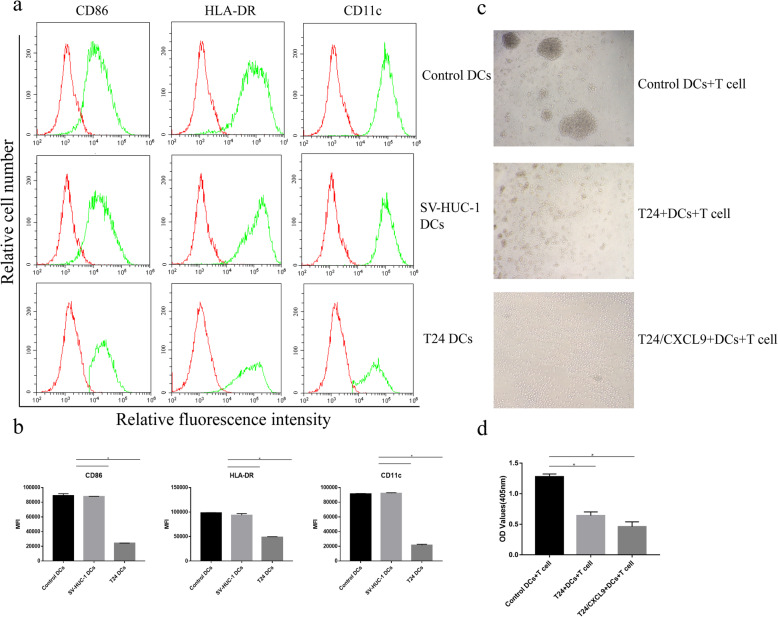
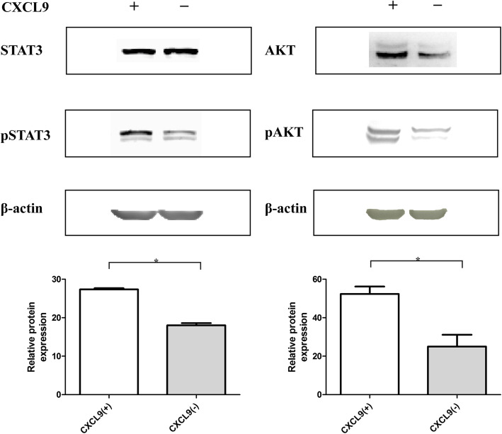
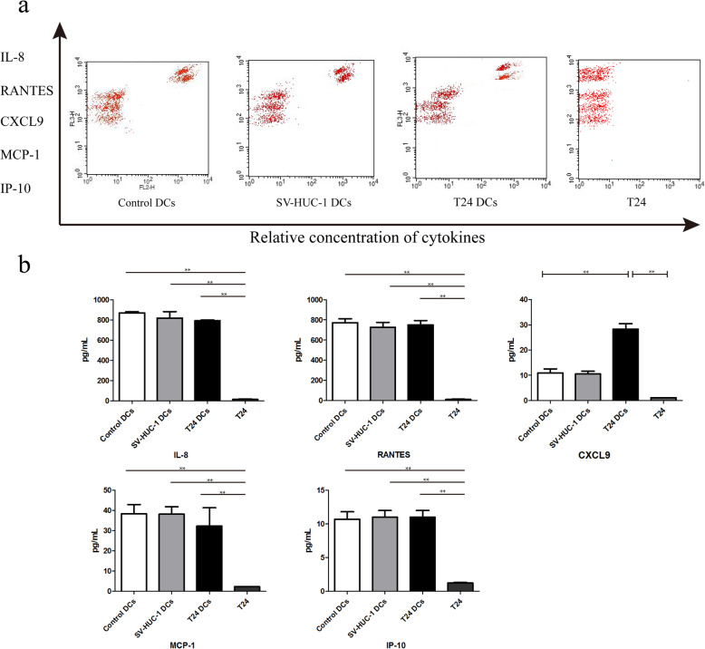
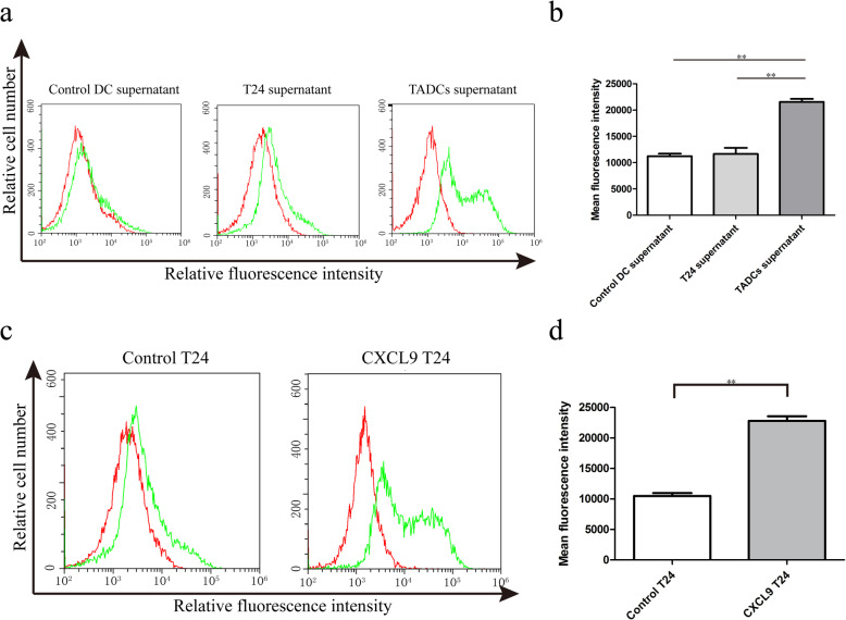
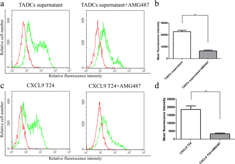
Compared with the control DCs, TADCs exhibited immature phenotype and had significantly lower capacity to stimulate allogenic T cell proliferation, particularly in the presence of recombinant CXCL9. TADCs produced significantly higher levels of CXCL9, which enhanced PD-L1 expression in T24 cells. Pre-treatment with AMG487 abrogated the CXCL9-increased PD-L1 expression in T24 cells. Treatment with CXCL9 significantly enhanced STAT3 and Akt activation in T24 cells.
TADCs produced high levels of CXCL9 that increased PD-L1 expression in bladder cancer T24 cells by activating the CXCR3-related signaling. Our findings may shed new lights in understanding the regulatory roles of TADCs in inhibiting antitumor T cell responses and promoting tumor growth.
肿瘤相关树突状细胞(TADCs)可与肿瘤细胞相互作用,抑制抗肿瘤 T 细胞免疫。然而,目前尚不清楚 TADCs 是否以及如何调节癌细胞程序性死亡配体 1(PD-L1)的表达。
人外周血单核细胞被诱导为树突状细胞,不成熟的树突状细胞单独培养或与膀胱癌 T24 或对照 SV-HUC-1 细胞共培养,然后用 LPS 刺激树突状细胞活化。通过流式细胞术和同种异体 T 细胞增殖来表征树突状细胞的活化状态。通过基于 CBA 的流式细胞术测量共培养树突状细胞上清液中趋化因子的水平。通过流式细胞术和 Western blot 确定 CXCL9 对 T24 细胞中 PD-L1、STAT3 和 Akt 表达以及 STAT3 和 Akt 磷酸化的影响。
与对照树突状细胞相比,TADCs 表现出不成熟的表型,刺激同种异体 T 细胞增殖的能力显著降低,尤其是在存在重组 CXCL9 的情况下。TADCs 产生的 CXCL9 水平明显更高,这增强了 T24 细胞中的 PD-L1 表达。用 AMG487 预处理可消除 CXCL9 对 T24 细胞中 PD-L1 表达的增加。CXCL9 处理显著增强了 T24 细胞中 STAT3 和 Akt 的激活。
TADCs 产生高水平的 CXCL9,通过激活 CXCR3 相关信号通路,增加膀胱癌 T24 细胞中 PD-L1 的表达。我们的研究结果可能为理解 TADCs 在抑制抗肿瘤 T 细胞反应和促进肿瘤生长中的调节作用提供新的思路。