Department of General, Visceral, and Transplant Surgery, Ludwig-Maximilians-University Munich, Munich, Germany.
German Cancer Consortium (DKTK), Partner Site Munich, Munich, Germany.
Front Immunol. 2018 Sep 27;9:2129. doi: 10.3389/fimmu.2018.02129. eCollection 2018.
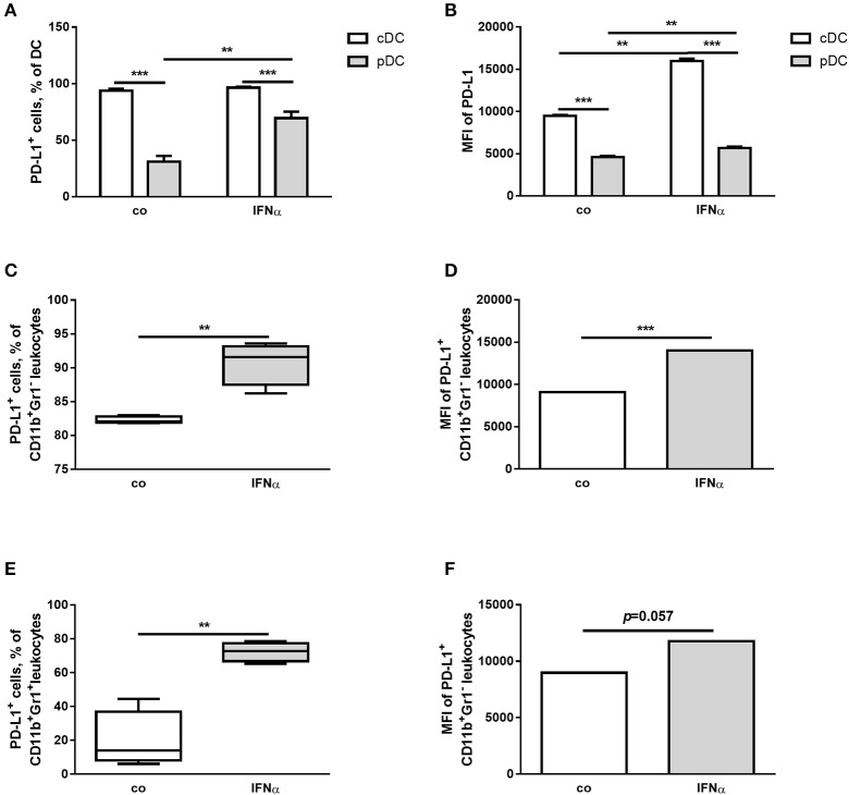
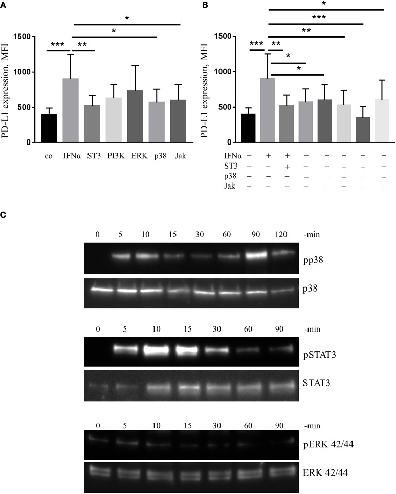
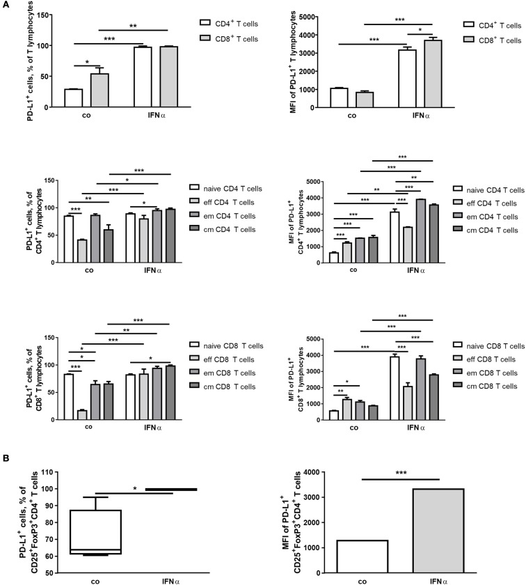
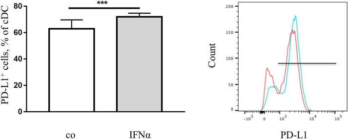
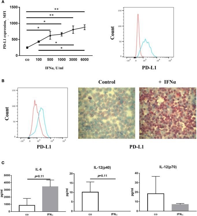
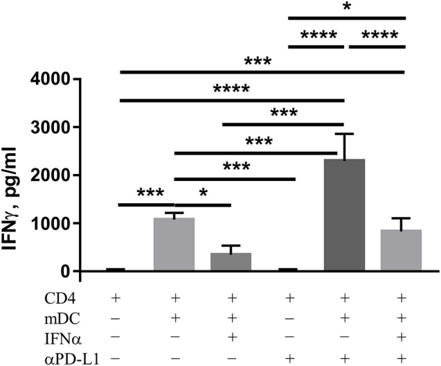
Interferon-α (IFNα) has one of the longest histories of use amongst cytokines in clinical oncology and has been applied for the treatment of many types of cancers. Due to its immune-activating properties, IFNα is also an attractive candidate for combinatory anti-cancer therapies. Despite its extensive use in animal tumor models as well as in several clinical trials, the different mechanisms underlying patient responses and affecting desirable clinical benefits are still under investigation. Here we show that in addition to its immune-activating properties, IFNα induces the expression of a key negative regulator, immunosuppressive PD-L1 molecule, in the majority of the specific immune cell populations, particularly in the dendritic cells (DC). DC can modulate immune responses by a variety of mechanisms, including expression of T-cell regulatory molecules and cytokines. Our results showed that treatment of DC with IFNα-2b led to pronounced up-regulation of surface expression of PD-L1 molecules, increased IL-6 and decreased IL-12 production. Moreover, we present evidence that IFNα-treated DC exhibited a reduced capacity to stimulate interferon-γ production in T cells compared to control DC. This T-cell response after treatment of DC with IFNα was recovered by a pre-treatment with an anti-PD-L1 blocking antibody. Further analyses revealed that IFNα regulated PD-L1 expression through the STAT3 and p38 signaling pathways, since blocking of STAT3 and p38 activation with specific inhibitors prevented PD-L1 up-regulation. Our findings underline the important roles of p38 and STAT3 in the regulation of PD-L1 expression and prove that IFNα induces STAT3/p38-mediated expression of PD-L1 and thereby a reduced stimulatory ability of DC. The augmentation of PD-L1 expression in immune cells through IFNα treatment should be considered by use of IFNα in an anti-cancer therapy.
干扰素-α(IFNα)是在临床肿瘤学中应用时间最长的细胞因子之一,已用于治疗多种癌症。由于其免疫激活特性,IFNα也是联合抗癌治疗的有吸引力的候选药物。尽管它在动物肿瘤模型以及几项临床试验中得到了广泛应用,但仍在研究导致患者反应不同和影响理想临床获益的不同机制。在这里,我们表明,除了其免疫激活特性外,IFNα还会诱导大多数特定免疫细胞群体(尤其是树突状细胞(DC))中关键负调控因子、免疫抑制性 PD-L1 分子的表达。DC 可以通过多种机制调节免疫反应,包括表达 T 细胞调节分子和细胞因子。我们的结果表明,IFNα-2b 处理 DC 会导致 PD-L1 分子表面表达明显上调,IL-6 增加和 IL-12 减少。此外,我们还提供了证据表明,与对照 DC 相比,IFNα 处理的 DC 刺激 T 细胞产生干扰素-γ的能力降低。用抗 PD-L1 阻断抗体预处理可恢复用 IFNα 处理 DC 后的 T 细胞反应。进一步的分析表明,IFNα 通过 STAT3 和 p38 信号通路调节 PD-L1 的表达,因为用特异性抑制剂阻断 STAT3 和 p38 激活可防止 PD-L1 的上调。我们的研究结果强调了 p38 和 STAT3 在 PD-L1 表达调控中的重要作用,并证明 IFNα 诱导 STAT3/p38 介导的 PD-L1 表达,从而降低 DC 的刺激能力。通过 IFNα 治疗增强免疫细胞中的 PD-L1 表达,在使用 IFNα 进行抗癌治疗时应予以考虑。