• 文献检索
  • 文档翻译
  • 深度研究
  • 学术资讯
  • Suppr Zotero 插件Zotero 插件
  • 邀请有礼
  • 套餐&价格
  • 历史记录
应用&插件
Suppr Zotero 插件Zotero 插件浏览器插件Mac 客户端Windows 客户端微信小程序
定价
高级版会员购买积分包购买API积分包
服务
文献检索文档翻译深度研究API 文档MCP 服务
关于我们
关于 Suppr公司介绍联系我们用户协议隐私条款
关注我们

Suppr 超能文献

核心技术专利:CN118964589B侵权必究
粤ICP备2023148730 号-1Suppr @ 2026

文献检索

告别复杂PubMed语法,用中文像聊天一样搜索,搜遍4000万医学文献。AI智能推荐,让科研检索更轻松。

立即免费搜索

文件翻译

保留排版,准确专业,支持PDF/Word/PPT等文件格式,支持 12+语言互译。

免费翻译文档

深度研究

AI帮你快速写综述,25分钟生成高质量综述,智能提取关键信息,辅助科研写作。

立即免费体验

MR-PWI 定量和半定量参数在肝纤维化评估中的比较。

Comparison of MR-PWI quantitative and semi-quantitative parameters for the evaluation of liver fibrosis.

机构信息

Department of Radiology, The Third Affiliated Hospital of Guangxi Medical University, No. 13, Dancun Road, Nanning, 530031, China.

Department of Ultrasound, The Third Affiliated Hospital of Guangxi Medical University, Nanning, 530031, China.

出版信息

BMC Med Imaging. 2021 Jan 6;21(1):8. doi: 10.1186/s12880-020-00539-3.

DOI:10.1186/s12880-020-00539-3
PMID:33407215
原文链接:https://pmc.ncbi.nlm.nih.gov/articles/PMC7789507/
Abstract

BACKGROUND

To evaluate different stages of liver fibrosis in cynomolgus monkeys by comparing magnetic resonance-perfusion weighted imaging (MR-PWI) quantitative and semi-quantitative parameters, and confirm the best detection indicators for diagnosis of liver fibrosis.

METHODS

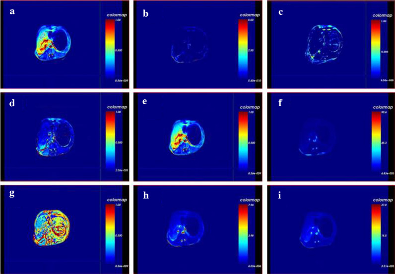
A liver fibrosis model of different stages (S0-S4) was established in cynomolgus monkeys. The changes in MR-PWI quantitative and semi-quantitative parameters with the progression of liver fibrosis were investigated.

RESULTS

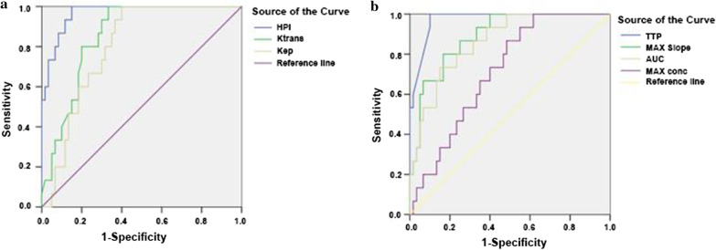
MR-PWI quantitative parameters gradually decreased with the progression of liver fibrosis. Hepatic arterial perfusion index (HPI) was found to increase with the progression of liver fibrosis and significant differences of HPI between each group were observed. There was a highly positive correlation between HPI and the stages of liver fibrosis. Receiver operating characteristic (ROC) curve analysis showed that HPI had the highest efficacy of the MR-PWI quantitative parameters for the diagnosis of liver fibrosis. The MR-PW semi-quantitative parameters gradually reduced with the progression of liver fibrosis, and the differences were statistically significant between stages S3-S4 and S0-S2. Time to peak (TPP) gradually extended and showed a positive correlation with the stages of liver fibrosis. TTP had the highest efficacy of the semi-quantitative parameters for diagnosis of liver fibrosis.

CONCLUSIONS

Both the MR-PWI quantitative and semi-quantitative parameters of the liver fibrosis model in cynomolgus monkeys varied at different stages of liver fibrosis, and HPI and TTP were the best detection indices for quantitative and semi-quantitative evaluation of liver fibrosis, respectively.

摘要

背景

通过比较磁共振灌注加权成像(MR-PWI)定量和半定量参数,评估食蟹猴不同阶段的肝纤维化,并确定最佳检测指标用于诊断肝纤维化。

方法

建立食蟹猴不同阶段(S0-S4)的肝纤维化模型,研究 MR-PWI 定量和半定量参数随肝纤维化进展的变化。

结果

MR-PWI 定量参数随肝纤维化的进展逐渐降低。肝动脉灌注指数(HPI)随肝纤维化的进展而升高,各组之间 HPI 差异有统计学意义。HPI 与肝纤维化分期呈高度正相关。受试者工作特征(ROC)曲线分析显示,HPI 对肝纤维化的诊断具有最高的 MR-PWI 定量参数效能。MR-PW 半定量参数随肝纤维化的进展逐渐降低,分期 S3-S4 与 S0-S2 之间差异有统计学意义。达峰时间(TPP)逐渐延长,与肝纤维化分期呈正相关。TPP 对半定量参数诊断肝纤维化的效能最高。

结论

食蟹猴肝纤维化模型的 MR-PWI 定量和半定量参数在肝纤维化的不同阶段均有变化,HPI 和 TPP 分别是定量和半定量评估肝纤维化的最佳检测指标。

https://cdn.ncbi.nlm.nih.gov/pmc/blobs/1479/7789507/553a660cbb9a/12880_2020_539_Fig3_HTML.jpg
https://cdn.ncbi.nlm.nih.gov/pmc/blobs/1479/7789507/c511fc9b6fa4/12880_2020_539_Fig1_HTML.jpg
https://cdn.ncbi.nlm.nih.gov/pmc/blobs/1479/7789507/fca4bd3309fd/12880_2020_539_Fig2_HTML.jpg
https://cdn.ncbi.nlm.nih.gov/pmc/blobs/1479/7789507/553a660cbb9a/12880_2020_539_Fig3_HTML.jpg
https://cdn.ncbi.nlm.nih.gov/pmc/blobs/1479/7789507/c511fc9b6fa4/12880_2020_539_Fig1_HTML.jpg
https://cdn.ncbi.nlm.nih.gov/pmc/blobs/1479/7789507/fca4bd3309fd/12880_2020_539_Fig2_HTML.jpg
https://cdn.ncbi.nlm.nih.gov/pmc/blobs/1479/7789507/553a660cbb9a/12880_2020_539_Fig3_HTML.jpg

相似文献

1
Comparison of MR-PWI quantitative and semi-quantitative parameters for the evaluation of liver fibrosis.MR-PWI 定量和半定量参数在肝纤维化评估中的比较。
BMC Med Imaging. 2021 Jan 6;21(1):8. doi: 10.1186/s12880-020-00539-3.
2
[A comparative study of 3.0T (1)H-MRS for varying degrees of liver fibrosis in cynomolgus monkeys].食蟹猴不同程度肝纤维化的3.0T氢质子磁共振波谱对比研究
Zhonghua Gan Zang Bing Za Zhi. 2020 Sep 20;28(9):766-772. doi: 10.3760/cma.j.cn501113-20190212-00044.
3
[Establishment of liver fibrosis in rabbit model and quantitative study on hepatic perfusion with dynamic whole-liver 3D MR imaging].[兔肝纤维化模型的建立及动态全肝三维磁共振成像对肝脏灌注的定量研究]
Zhonghua Gan Zang Bing Za Zhi. 2009 May;17(5):350-3.
4
Contrast-enhanced ultrasound and computerized tomography perfusion imaging of a liver fibrosis-early cirrhosis in dogs.犬肝纤维化-早期肝硬化的超声造影及计算机断层扫描灌注成像
J Gastroenterol Hepatol. 2016 Sep;31(9):1604-10. doi: 10.1111/jgh.13320.
5
Application Value of Magnetic Resonance Perfusion Imaging in the Early Diagnosis of Rat Hepatic Fibrosis.磁共振灌注成像在大鼠肝纤维化早期诊断中的应用价值。
Biomed Res Int. 2019 Dec 28;2019:5095934. doi: 10.1155/2019/5095934. eCollection 2019.
6
Liver extracellular volume fraction values obtained with magnetic resonance imaging can quantitatively stage liver fibrosis: a validation study in monkeys with nonalcoholic steatohepatitis.磁共振成像获取的肝细胞外容积分数值可定量分期肝纤维化:非酒精性脂肪性肝炎猴模型的验证研究。
Eur Radiol. 2020 Oct;30(10):5748-5757. doi: 10.1007/s00330-020-06902-w. Epub 2020 May 6.
7
Intrahepatic transit time predicts liver fibrosis in patients with chronic hepatitis B: quantitative assessment with contrast-enhanced ultrasonography.超声造影定量分析肝内超声时间预测慢性乙型肝炎肝纤维化
Ultrasound Med Biol. 2010 Jul;36(7):1066-75. doi: 10.1016/j.ultrasmedbio.2010.04.012.
8
Quantification of Hemodynamic Changes in Chronic Liver Disease: Correlation of Perfusion-CT Data with Histopathologic Staging of Fibrosis.慢性肝脏疾病血流动力学变化的定量分析:灌注 CT 数据与纤维化组织病理分期的相关性。
Acad Radiol. 2019 Sep;26(9):1174-1180. doi: 10.1016/j.acra.2018.11.009. Epub 2018 Dec 6.
9
3D-liver perfusion MRI with the MS-325 blood pool agent: a noninvasive protocol to asses liver fibrosis.采用 MS-325 血池造影剂的 3D-肝脏灌注 MRI:一种评估肝纤维化的非侵入性方案。
J Magn Reson Imaging. 2012 Jun;35(6):1380-7. doi: 10.1002/jmri.23594. Epub 2012 Jan 26.
10
A study of free portal pressure in cynomolgus monkeys with different degrees of liver fibrosis.食蟹猴不同程度肝纤维化时门静脉自由压的研究。
J Environ Pathol Toxicol Oncol. 2014;33(4):315-21. doi: 10.1615/jenvironpatholtoxicoloncol.2014011492.

引用本文的文献

1
Hepatic microcirculatory disturbance in liver diseases: intervention with traditional Chinese medicine.肝脏疾病中的肝微循环障碍:中医药干预
Front Pharmacol. 2024 Jul 23;15:1399598. doi: 10.3389/fphar.2024.1399598. eCollection 2024.
2
Neuronal Apoptosis in Patients with Liver Cirrhosis and Neuronal Epileptiform Discharge Model Based upon Multi-Modal Fusion Deep Learning.基于多模态融合深度学习的肝硬化患者神经元凋亡和神经元癫痫样放电模型。
J Healthc Eng. 2022 Mar 17;2022:2203737. doi: 10.1155/2022/2203737. eCollection 2022.

本文引用的文献

1
Application Value of Magnetic Resonance Perfusion Imaging in the Early Diagnosis of Rat Hepatic Fibrosis.磁共振灌注成像在大鼠肝纤维化早期诊断中的应用价值。
Biomed Res Int. 2019 Dec 28;2019:5095934. doi: 10.1155/2019/5095934. eCollection 2019.
2
siRNA- and miRNA-based therapeutics for liver fibrosis.基于 siRNA 和 miRNA 的肝纤维化治疗方法。
Transl Res. 2019 Dec;214:17-29. doi: 10.1016/j.trsl.2019.07.007. Epub 2019 Aug 13.
3
Liver fibrosis imaging: A clinical review of ultrasound and magnetic resonance elastography.肝脏纤维化成像:超声和磁共振弹性成像的临床综述。
J Magn Reson Imaging. 2020 Jan;51(1):25-42. doi: 10.1002/jmri.26716. Epub 2019 Mar 12.
4
SIRT6 deficiency results in developmental retardation in cynomolgus monkeys.SIRT6 缺乏导致食蟹猴发育迟缓。
Nature. 2018 Aug;560(7720):661-665. doi: 10.1038/s41586-018-0437-z. Epub 2018 Aug 22.
5
Magnetic Resonance Diffusion Kurtosis Imaging for Evaluating Stage of Liver Fibrosis in a Rabbit Model.磁共振扩散峰度成像评价兔肝纤维化分期的价值。
Acad Radiol. 2019 Jun;26(6):e90-e97. doi: 10.1016/j.acra.2018.06.018. Epub 2018 Jul 31.
6
Liver fibrosis: Direct antifibrotic agents and targeted therapies.肝纤维化:直接抗纤维化药物和靶向治疗。
Matrix Biol. 2018 Aug;68-69:435-451. doi: 10.1016/j.matbio.2018.04.006. Epub 2018 Apr 12.
7
Quantitative and noninvasive assessment of chronic liver diseases using two-dimensional shear wave elastography.应用二维剪切波弹性成像技术对慢性肝脏疾病进行定量及无创评估。
World J Gastroenterol. 2018 Mar 7;24(9):957-970. doi: 10.3748/wjg.v24.i9.957.
8
Prospects in non-invasive assessment of liver fibrosis: Liquid biopsy as the future gold standard?肝纤维化无创评估的前景:液体活检是否会成为未来的金标准?
Biochim Biophys Acta Mol Basis Dis. 2018 Apr;1864(4 Pt A):1024-1036. doi: 10.1016/j.bbadis.2018.01.009. Epub 2018 Jan 9.
9
Diffusion kurtosis imaging with the breath-hold technique for staging hepatic fibrosis: A preliminary study.屏气技术扩散峰度成像用于肝纤维化分期的初步研究
Magn Reson Imaging. 2018 Apr;47:33-38. doi: 10.1016/j.mri.2017.11.001. Epub 2017 Nov 20.
10
Shear wave liver elastography.剪切波肝脏弹性成像。
Abdom Radiol (NY). 2018 Apr;43(4):800-807. doi: 10.1007/s00261-017-1375-1.