Demirel Pinar B, Dogan Soner, Ozorhan Umit, Tuna Bilge G, Schuster Todd F, Cleary Margot P
Department of Medical Biology and Genetics, Faculty of Medicine, Maltepe University, Istanbul, Turkey.
Department of Medical Biology, Faculty of Medicine, Yeditepe University, Istanbul, Turkey.
J Exp Clin Med (Samsun). 2020;37(4):119-125. Epub 2020 Nov 9.
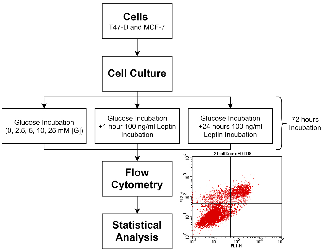
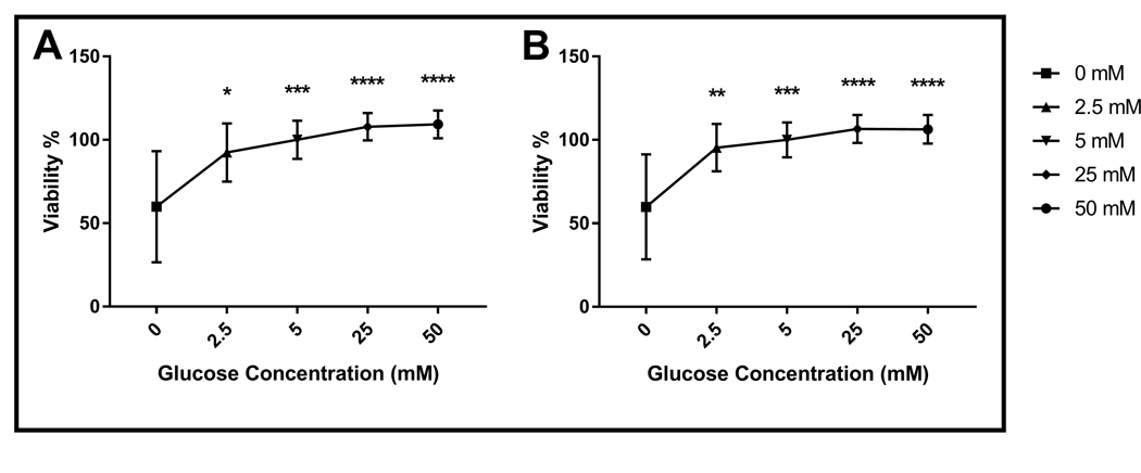
Obesity is associated with increased risk of breast cancer. Leptin is a well-known factor involved in obesity and its serum levels are increased in breast cancer. Hyperglycemia is another significant risk factor for breast cancer. Consistently, high glucose induces proliferation and invasion of breast cancer cells and in-vivo calorie restriction reduce tumorigenesis in rodent models. The aim of this study was to investigate the effect of leptin on the viability and mode of cell death in breast cancer cells incubated in different glucose concentrations to represent caloric restriction. For this purpose, MCF-7 and T47D breast cancer cells incubated in different glucose concentrations for a total of 72 hours were treated with or without leptin either for one hour or 24 hours and the ratio of apoptotic, necrotic and alive cells were analyzed by flow cytometry. Our data revealed that glucose incubation significantly decreased apoptosis and necrosis, while increasing viability in both cell lines in a dose dependent manner. One-hour leptin treatment significantly decreased viability, and increased apoptosis but did not significantly affect necrosis in T47D cells incubated in 2.5 mM glucose. In MCF-7 cells, one-hour leptin incubation significantly increased necrosis but its effects on apoptosis and viability were not significant. In conclusion, although glucose induces cell death by apoptosis and necrosis in T47D and MCF-7 cells respectively in a dose dependent manner, the overallviability is still increased in both cell lines. One-hour leptin treatment reverses the effect of low glucose incubation on apoptosis of T47D and necrosis of MCF-7 cells. Moreover, the effect of one-hour leptin treatment on apoptosis or necrosis is significantly higher than that of 24-hour leptin treatment.
肥胖与乳腺癌风险增加相关。瘦素是一种与肥胖有关的知名因子,其血清水平在乳腺癌中会升高。高血糖是乳腺癌的另一个重要风险因素。一致的是,高糖会诱导乳腺癌细胞的增殖和侵袭,而在啮齿动物模型中,体内热量限制可减少肿瘤发生。本研究的目的是研究瘦素对在不同葡萄糖浓度下培养以模拟热量限制的乳腺癌细胞活力和细胞死亡方式的影响。为此,将MCF-7和T47D乳腺癌细胞在不同葡萄糖浓度下共培养72小时,分别用或不用瘦素处理1小时或24小时,然后通过流式细胞术分析凋亡、坏死和存活细胞的比例。我们的数据显示,葡萄糖培养显著降低了凋亡和坏死,同时以剂量依赖的方式增加了两种细胞系的活力。在2.5 mM葡萄糖中培养的T47D细胞中,1小时的瘦素处理显著降低了活力,增加了凋亡,但对坏死没有显著影响。在MCF-7细胞中,1小时的瘦素培养显著增加了坏死,但其对凋亡和活力的影响不显著。总之,尽管葡萄糖分别以剂量依赖的方式在T47D和MCF-7细胞中通过凋亡和坏死诱导细胞死亡,但两种细胞系的总体活力仍然增加。1小时的瘦素处理逆转了低葡萄糖培养对T47D细胞凋亡和MCF-7细胞坏死的影响。此外,1小时瘦素处理对凋亡或坏死的影响显著高于24小时瘦素处理。